Deposition Date
2009-07-24
Release Date
2010-07-28
Last Version Date
2023-11-01
Entry Detail
PDB ID:
3A51
Keywords:
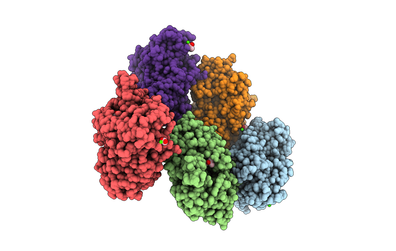
Title:
Structure of cytochrome P450 Vdh mutant (Vdh-K1) obtained by directed evolution with bound 25-hydroxyvitamin D3
Biological Source:
Source Organism(s):
Pseudonocardia autotrophica (Taxon ID: 2074)
Expression System(s):
Method Details:
Experimental Method:
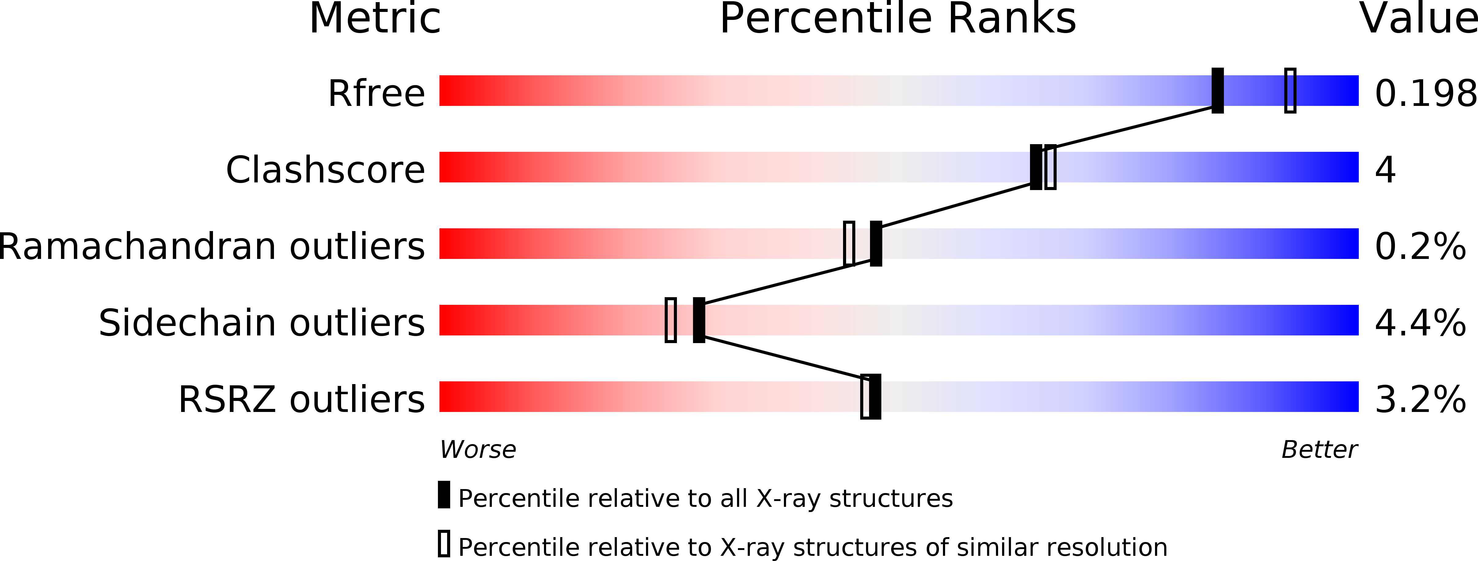
Resolution:
2.00 Å
R-Value Free:
0.23
R-Value Work:
0.19
R-Value Observed:
0.19
Space Group:
P 21 21 21