Deposition Date
2009-06-23
Release Date
2009-09-08
Last Version Date
2024-11-13
Entry Detail
PDB ID:
3A3Y
Keywords:
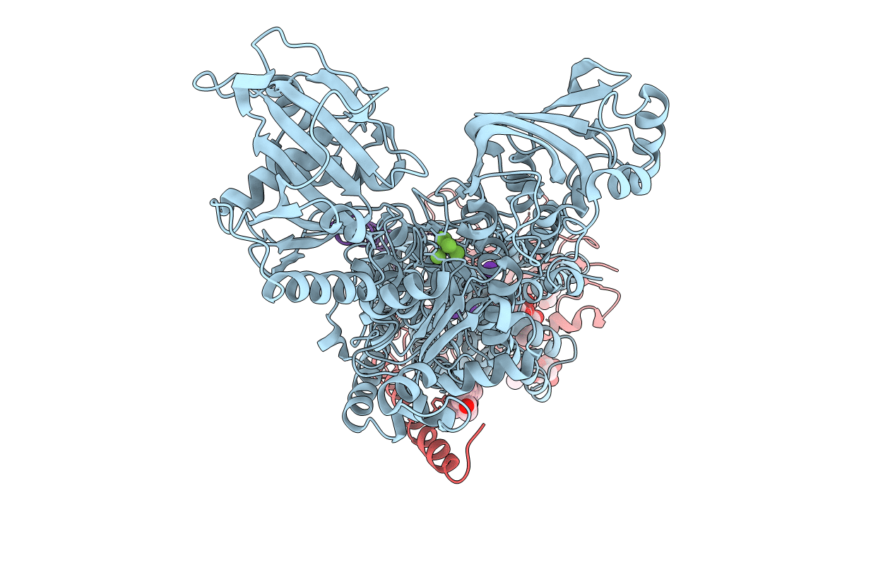
Title:
Crystal structure of the sodium-potassium pump with bound potassium and ouabain
Biological Source:
Source Organism(s):
Squalus acanthias (Taxon ID: 7797)
Method Details:
Experimental Method:
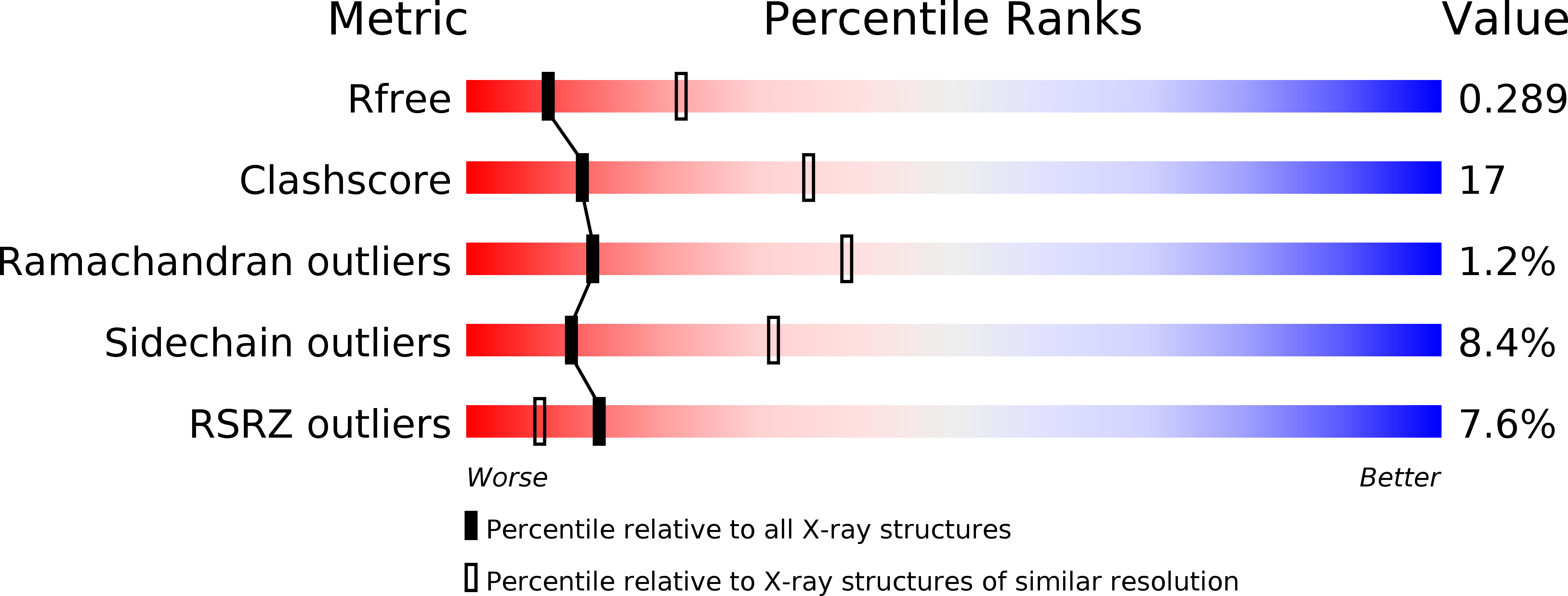
Resolution:
2.80 Å
R-Value Free:
0.29
R-Value Work:
0.24
R-Value Observed:
0.24
Space Group:
C 1 2 1