3A37 image
Deposition Date 2009-06-10
Release Date 2010-01-12
Last Version Date 2024-03-13
Entry Detail
PDB ID:
3A37
Keywords:
Title:
Structural insight into the membrane insertion of tail-anchored proteins by Get3
Biological Source:
Source Organism:
Host Organism:
Method Details:
Experimental Method:
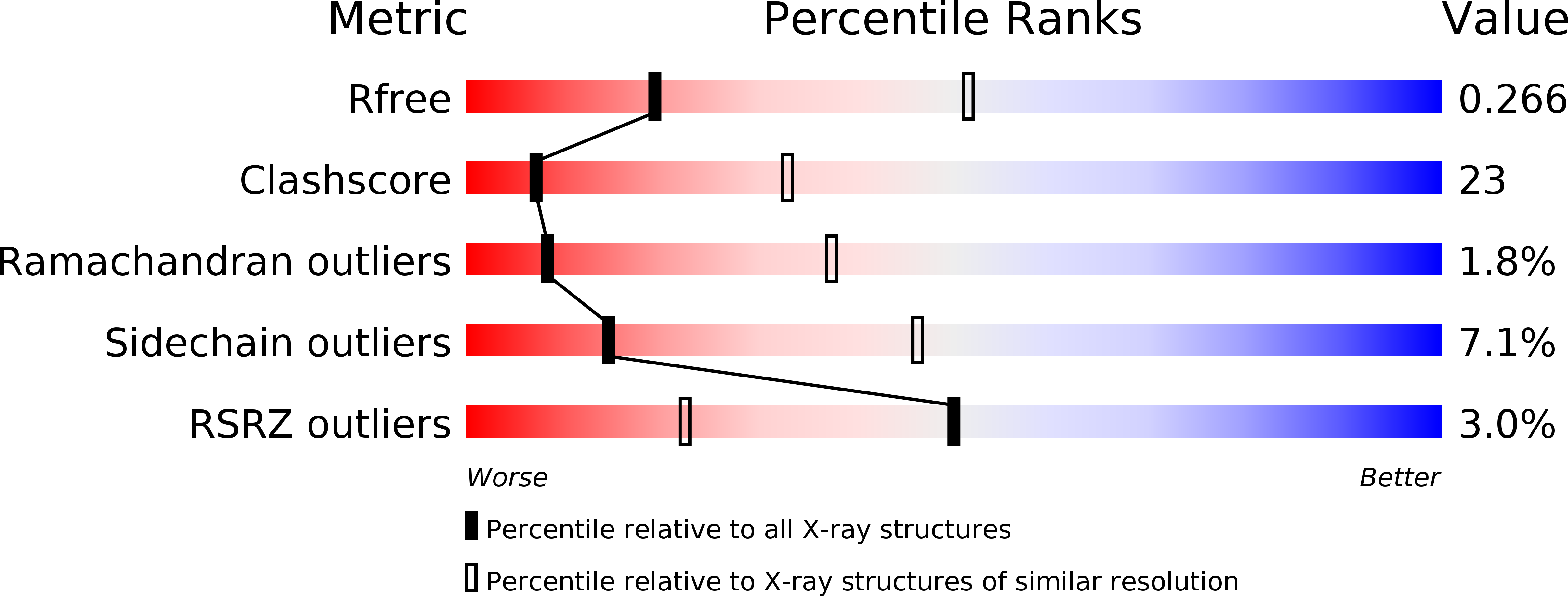
Resolution:
3.00 Å
R-Value Free:
0.28
R-Value Work:
0.25
R-Value Observed:
0.25
Space Group:
P 21 21 2
Macromolecular Entities
Polymer Type:polypeptide(L)
Molecule:ATPase GET3
Gene (Uniprot):GET3
Chain IDs:A, B
Chain Length:362
Number of Molecules:2
Biological Source:Saccharomyces cerevisiae
Primary Citation
Structural insight into the membrane insertion of tail-anchored proteins by Get3
Genes Cells 15 29 41 (2010)
PMID: 20015340 DOI: 10.1111/j.1365-2443.2009.01362.x

Abstact

Tail anchored (TA) proteins, which are important for numerous cellular processes, are defined by a single transmembrane domain (TMD) near the C-terminus. The membrane insertion of TA proteins is mediated by the highly conserved ATPase Get3. Here we report the crystal structures of Get3 in ADP-bound and nucleotide-free forms at 3.0 A and 2.8 A resolutions, respectively. Get3 consists of a nucleotide binding domain and a helical domain. Both structures exhibit a Zn(2+)-mediated homodimer in a head-to-head orientation, representing an open dimer conformation. Our cross-link experiments indicated the closed dimer-stimulating ATP hydrolysis, which might be coupled with TA-protein release. Further, our coexpression-based binding assays using a model TA protein Sec22p revealed the direct interaction between the helical domain of Get3 and the Sec22p TMD. This interaction is independent of ATP and dimer formation. Finally, we propose a structural mechanism that links ATP hydrolysis with the TA-protein insertion mediated by the conserved DTAPTGH motif.

Legend

Protein

Chemical

Disease

Primary Citation of related structures