3A2A image
Deposition Date 2009-05-08
Release Date 2010-02-09
Last Version Date 2024-10-23
Entry Detail
PDB ID:
3A2A
Title:
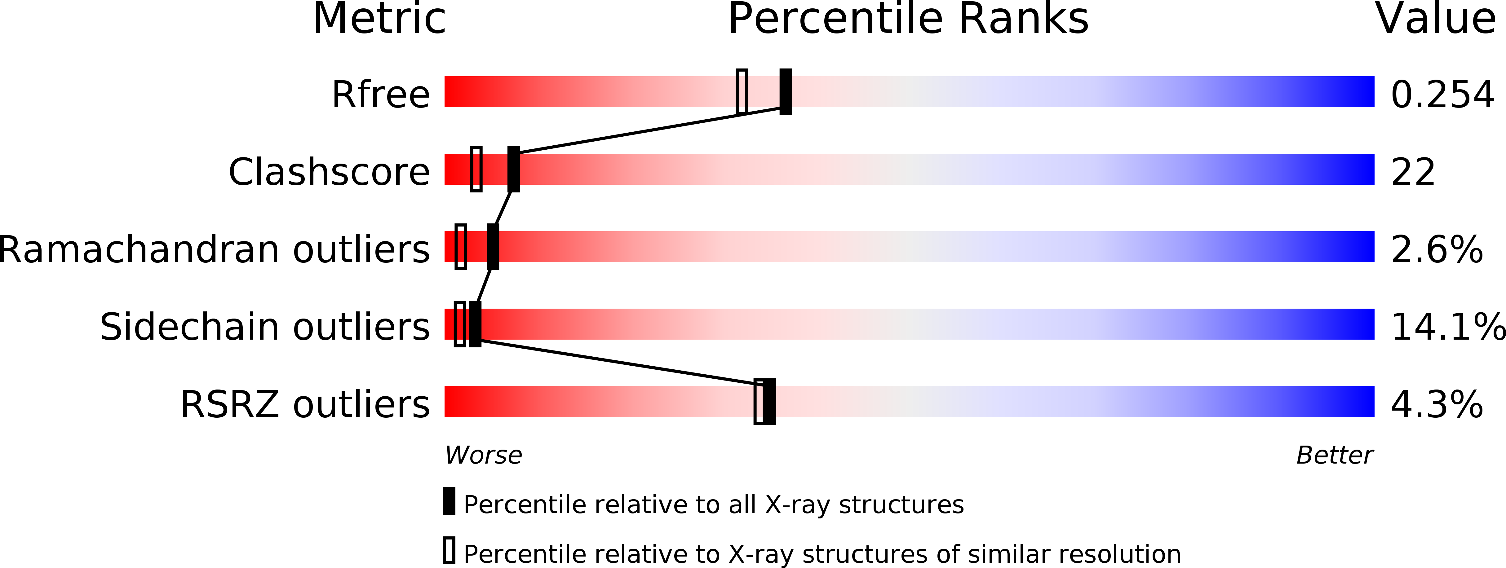
The structure of the carboxyl-terminal domain of the human voltage-gated proton channel Hv1
Biological Source:
Source Organism:
Homo sapiens (Taxon ID: 9606)
Host Organism:
Method Details:
Experimental Method:
Resolution:
2.00 Å
R-Value Free:
0.26
R-Value Work:
0.25
R-Value Observed:
0.25
Space Group:
P 43
Macromolecular Entities
Structures with similar UniProt ID
Protein Blast
Polymer Type:polypeptide(L)
Molecule:Voltage-gated hydrogen channel 1
Gene (Uniprot):HVCN1
Chain IDs:A, B, C, D
Chain Length:58
Number of Molecules:4
Biological Source:Homo sapiens
Ligand Molecules
Primary Citation
The role and structure of the carboxyl-terminal domain of the human voltage-gated proton channel Hv1.
J.Biol.Chem. 285 12047 12054 (2010)
PMID: 20147290 DOI: 10.1074/jbc.M109.040360

Abstact

The voltage-gated proton channel Hv1 has a voltage sensor domain but lacks a pore domain. Although the C-terminal domain of Hv1 is known to be responsible for dimeric architecture of the channel, its role and structure are not known. We report that the full-length Hv1 is mainly localized in intracellular compartment membranes rather than the plasma membrane. Truncation of either the N or C terminus alone or both together revealed that the N-terminal deletion did not alter localization, but deletion of the C terminus either alone or together with the N terminus resulted in expression throughout the cell. These results indicate that the C terminus is essential for Hv1 localization but not the N terminus. In the 2.0 A structure of the C-terminal domain, the two monomers form a dimer via a parallel alpha-helical coiled-coil, in which one chloride ion binds with the Neta atom of Arg(264). A pH-dependent structural change of the protein has been observed, but it remains a dimer irrespective of pH value.

Legend

Protein

Chemical

Disease

Primary Citation of related structures