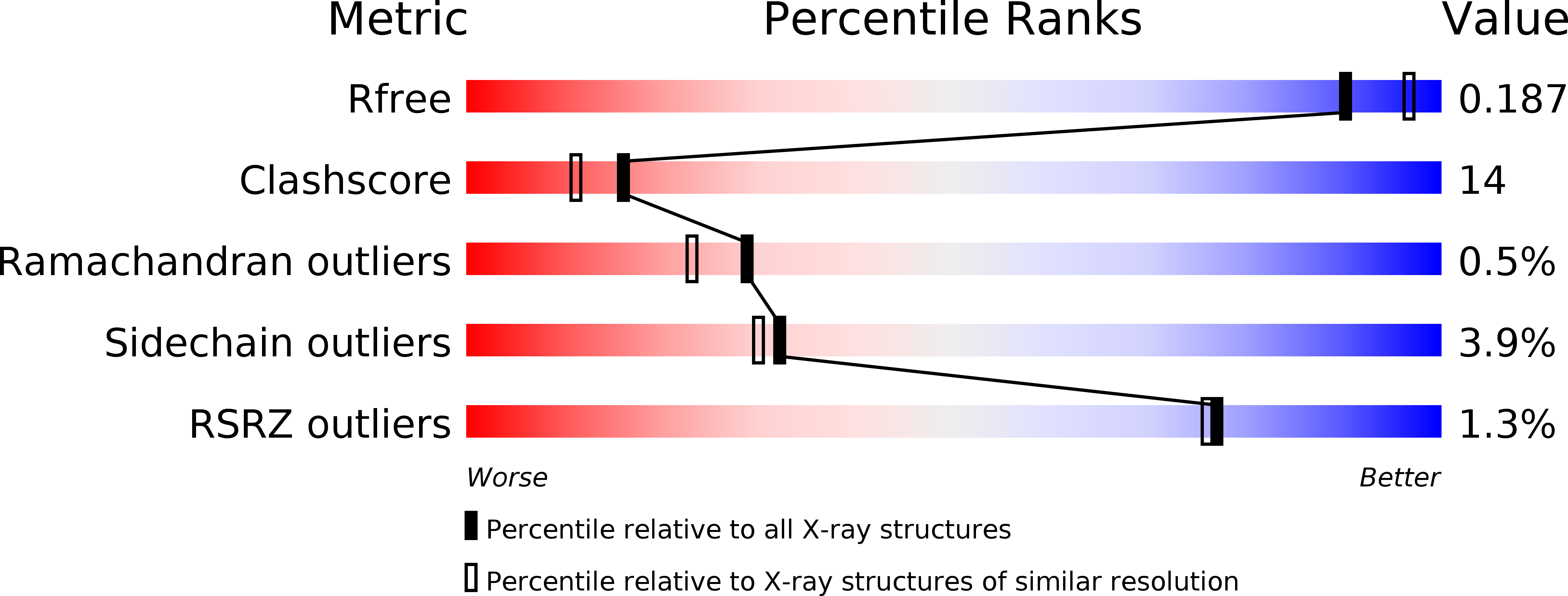
3A28 image
Deposition Date 2009-05-02
Release Date 2009-12-15
Last Version Date 2023-11-01
Entry Detail
PDB ID:
3A28
Keywords:
Title:
Crystal structure of L-2,3-butanediol dehydrogenase
Biological Source:
Source Organism:
Host Organism:
Method Details:
Experimental Method:
Resolution:
2.00 Å
R-Value Free:
0.24
R-Value Work:
0.19
Space Group:
P 1
Macromolecular Entities
Structures with similar UniProt ID
Protein Blast
Polymer Type:polypeptide(L)
Molecule:L-2.3-butanediol dehydrogenase
Gene (Uniprot):budC
Chain IDs:A (auth: C), B (auth: D), C (auth: A), D (auth: B), E, F, G, H
Chain Length:258
Number of Molecules:8
Biological Source:Brevibacterium saccharolyticum
Primary Citation
Structural basis for chiral substrate recognition by two 2,3-butanediol dehydrogenases
Febs Lett. 584 219 223 (2010)
PMID: 19941855 DOI: 10.1016/j.febslet.2009.11.068

Abstact

2,3-butanediol dehydrogenase (BDH) catalyzes the NAD-dependent redox reaction between acetoin and 2,3-butanediol. There are three types of homologous BDH, each stereospecific for both substrate and product. To establish how these homologous enzymes possess differential stereospecificities, we determined the crystal structure of l-BDH with a bound inhibitor at 2.0 A. Comparison with the inhibitor binding mode of meso-BDH highlights the role of a hydrogen-bond from a conserved Trp residue(192). Site-directed mutagenesis of three active site residues of meso-BDH, including Trp(190), which corresponds to Trp(192) of L-BDH, converted its stereospecificity to that of L-BDH. This result confirms the importance of conserved residues in modifying the stereospecificity of homologous enzymes.

Legend

Protein

Chemical

Disease

Primary Citation of related structures