3A14 image
Deposition Date 2009-03-25
Release Date 2010-04-07
Last Version Date 2024-03-13
Entry Detail
PDB ID:
3A14
Keywords:
Title:
Crystal structure of DXR from Thermotoga maritima, in complex with NADPH
Biological Source:
Source Organism:
Host Organism:
Method Details:
Experimental Method:
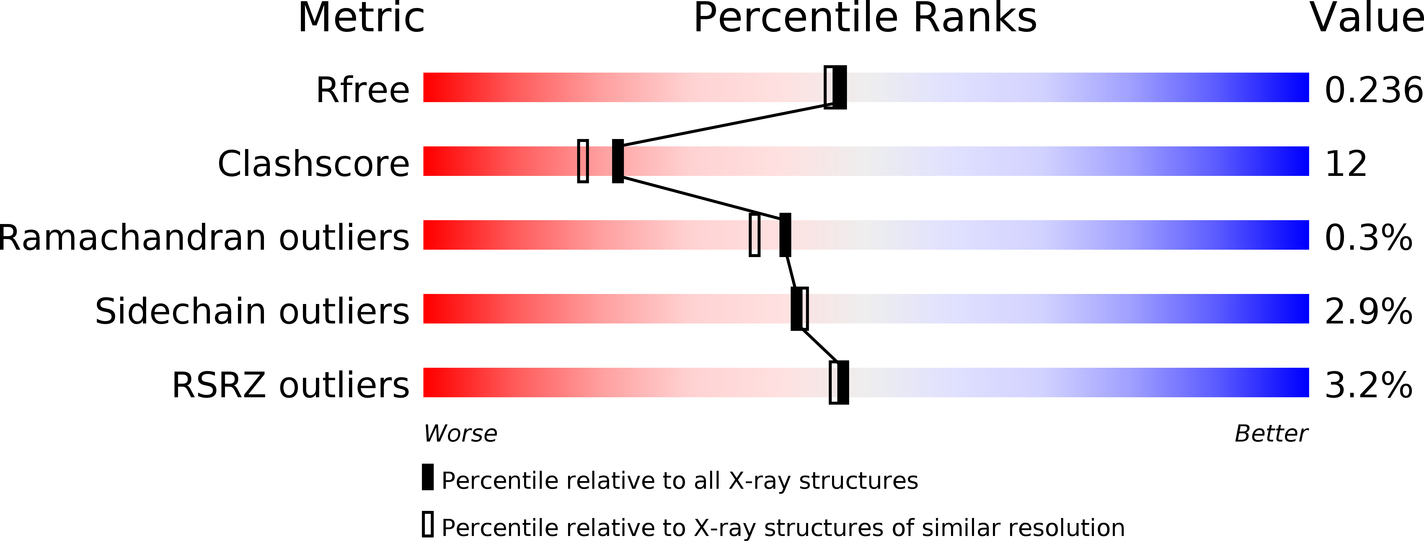
Resolution:
2.00 Å
R-Value Free:
0.23
R-Value Work:
0.20
R-Value Observed:
0.20
Space Group:
P 43
Macromolecular Entities
Structures with similar UniProt ID
Protein Blast
Polymer Type:polypeptide(L)
Molecule:1-deoxy-D-xylulose 5-phosphate reductoisomerase
Gene (Uniprot):dxr
Chain IDs:A, B
Chain Length:376
Number of Molecules:2
Biological Source:Thermotoga maritima
Primary Citation
Crystal structure of 1-deoxy-d-xylulose 5-phosphate reductoisomerase from the hyperthermophile Thermotoga maritima for insights into the coordination of conformational changes and an inhibitor binding.
J.Struct.Biol. ? ? ? (2010)
PMID: 20353826 DOI: 10.1016/j.jsb.2010.03.015

Abstact

Isopentenyl diphosphate is a precursor of various isoprenoids and is produced by the 2-C-methyl-d-erythritol 4-phosphate (MEP) pathway in plastids of plants, protozoa and many eubacteria. A key enzyme in the MEP pathway, 1-deoxy-d-xylulose 5-phosphate reductoisomerase (DXR), has been shown to be the target of fosmidomycin, which works as an antimalarial, antibacterial and herbicidal compound. In this paper, we report studies of kinetics and the crystal structures of the thermostable DXR from the hyperthermophile Thermotoga maritima. Unlike the mesophilic DXRs, Thermotoga DXR (tDXR) showed activity only with Mg(2+) at its growth temperature. We solved the crystal structures of tDXR with and without fosmidomycin. The structure without fosmidomycin but unexpectedly bound with 2-methyl-2,4-pentanediol (MPD), revealing a new extra space available for potential drug design. This structure adopted the closed form by rigid domain rotation but without the flexible loop over the active site, which was considered as a novel conformation. Further, the conserved Asp residue responsible for cation binding seemed to play an important role in adjusting the position of fosmidomycin. Taken together, our kinetic and the crystal structures illustrate the binding mode of fosmidomycin that leads to its slow, tight binding according to the conformational changes of DXR.

Legend

Protein

Chemical

Disease

Primary Citation of related structures