Planned Maintenance: Some services may turn out to be unavailable from 15th January, 2026 to 16th January, 2026. We apologize for the inconvenience!

3A0F image
Deposition Date 2009-03-16
Release Date 2009-09-08
Last Version Date 2024-10-16
Entry Detail
PDB ID:
3A0F
Keywords:
Title:
The crystal structure of Geotrichum sp. M128 xyloglucanase
Biological Source:
Source Organism(s):
Expression System(s):
Method Details:
Experimental Method:
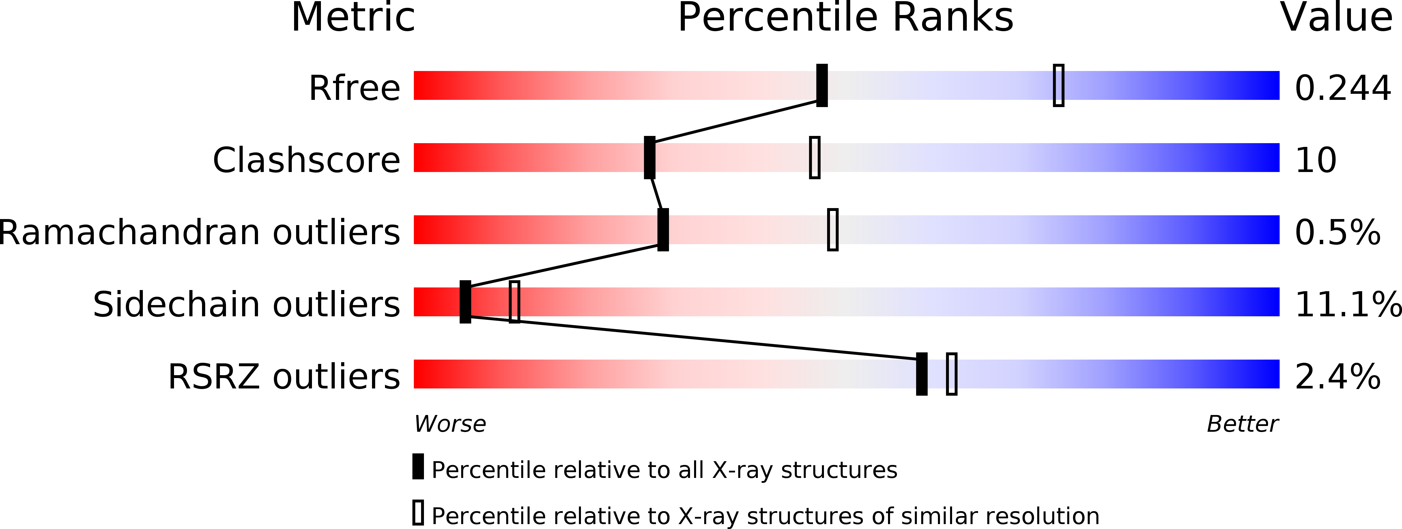
Resolution:
2.50 Å
R-Value Free:
0.27
R-Value Work:
0.23
R-Value Observed:
0.23
Space Group:
P 32 2 1
Macromolecular Entities
Structures with similar UniProt ID
Protein Blast
Polymer Type:polypeptide(L)
Molecule:Xyloglucanase
Chain IDs:A
Chain Length:763
Number of Molecules:1
Biological Source:Geotrichum sp. M128
Primary Citation
The crystal structure of a xyloglucan-specific endo-beta-1,4-glucanase from Geotrichum sp. M128 xyloglucanase reveals a key amino acid residue for substrate specificity
Febs J. 276 5094 5100 (2009)
PMID: 19682300 DOI: 10.1111/j.1742-4658.2009.07205.x

Abstact

Geotrichum sp. M128 possesses two xyloglucan-specific glycoside hydrolases belonging to family 74, xyloglucan-specific endo-beta-1,4-glucanase (XEG) and oligoxyloglucan reducing-end-specific cellobiohydrolase (OXG-RCBH). Despite their similar amino acid sequences (48% identity), their modes of action and substrate specificities are distinct. XEG catalyzes the hydrolysis of xyloglucan polysaccharides in endo mode, while OXG-RCBH acts on xyloglucan oligosaccharides at the reducing end in exo mode. Here, we determined the crystal structure of XEG at 2.5 A resolution, and compared it to a previously determined structure of OXG-RCBH. For the most part, the amino acid residues that interact with substrate are conserved between the two enzymes. However, there are notable differences at subsite positions -1 and +2. OXG-RCBH has a loop around the +2 site that blocks one end of the active site cleft, which accounts for its exo mode of action. In contrast, XEG lacks a corresponding loop at this site, thereby allowing binding to the middle of the main chain of the substrate. At the -1 site in OXG-RCBH, Asn488 interacts with the xylose side chain of the substrate, whereas the -1 site is occupied by Tyr457 in XEG. To confirm the contribution of this residue to substrate specificity, Tyr457 was substituted by Gly in XEG. The wild-type XEG cleaved the oligoxyloglucan at a specific site; the Y457G variant cleaved the same substrate, but at various sites. Together, the absence of a loop in the cleft and the presence of bulky Tyr457 determine the substrate specificity of XEG.

Legend

Protein

Chemical

Disease

Primary Citation of related structures
Feedback Form
Name
Email
Institute
Feedback