3A0B image
Deposition Date 2009-03-16
Release Date 2009-05-19
Last Version Date 2024-10-23
Entry Detail
PDB ID:
3A0B
Title:
Crystal structure of Br-substituted Photosystem II complex
Biological Source:
Source Organism:
Method Details:
Experimental Method:
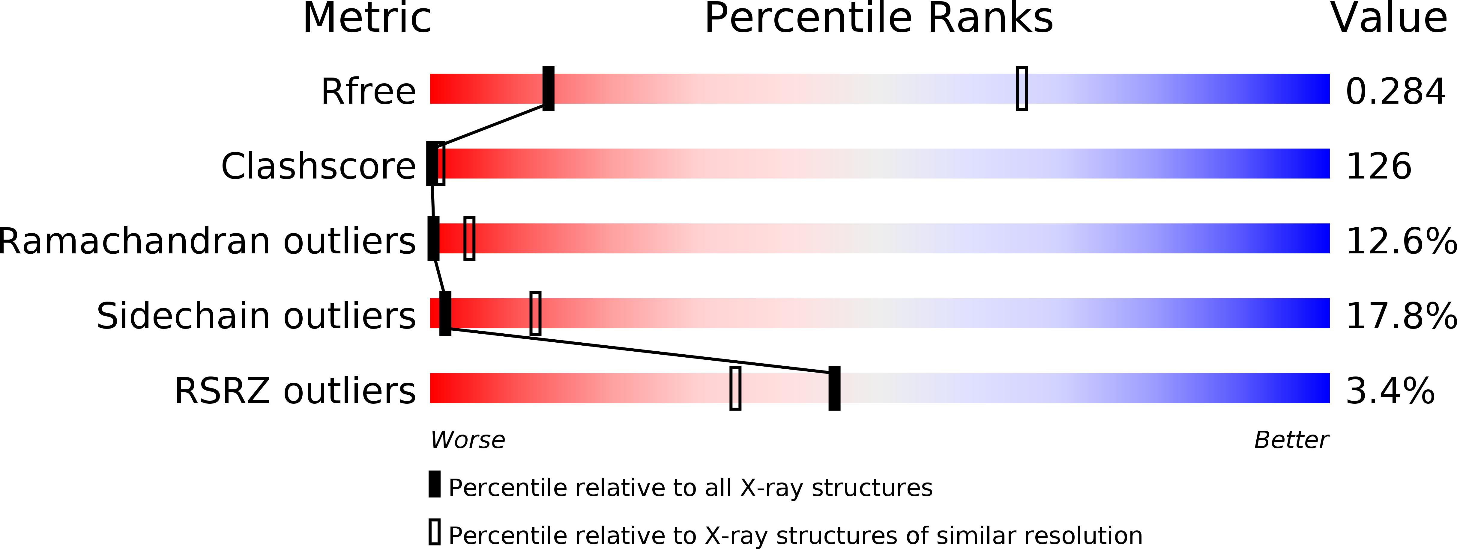
Resolution:
3.70 Å
R-Value Free:
0.35
R-Value Work:
0.30
R-Value Observed:
0.30
Space Group:
P 21 21 21
Macromolecular Entities
Structures with similar UniProt ID
Protein Blast
Polymer Type:polypeptide(L)
Molecule:Photosystem Q(B) protein
Gene (Uniprot):psbA
Chain IDs:A, U (auth: a)
Chain Length:344
Number of Molecules:2
Biological Source:Thermosynechococcus vulcanus
Structures with similar UniProt ID
Protein Blast
Polymer Type:polypeptide(L)
Molecule:Photosystem II core light harvesting protein
Chain IDs:B, V (auth: b)
Chain Length:488
Number of Molecules:2
Biological Source:Thermosynechococcus vulcanus
Structures with similar UniProt ID
Protein Blast
Polymer Type:polypeptide(L)
Molecule:Photosystem II CP43 protein
Chain IDs:C, W (auth: c)
Chain Length:447
Number of Molecules:2
Biological Source:Thermosynechococcus vulcanus
Structures with similar UniProt ID
Protein Blast
Polymer Type:polypeptide(L)
Molecule:Photosystem II D2 protein
Chain IDs:D, X (auth: d)
Chain Length:340
Number of Molecules:2
Biological Source:Thermosynechococcus vulcanus
Structures with similar UniProt ID
Protein Blast
Polymer Type:polypeptide(L)
Molecule:Cytochrome b559 subunit alpha
Gene (Uniprot):psbE
Chain IDs:E, Y (auth: e)
Chain Length:83
Number of Molecules:2
Biological Source:Thermosynechococcus vulcanus
Structures with similar UniProt ID
Protein Blast
Polymer Type:polypeptide(L)
Molecule:Cytochrome b559 subunit beta
Gene (Uniprot):psbF
Chain IDs:F, Z (auth: f)
Chain Length:44
Number of Molecules:2
Biological Source:Thermosynechococcus vulcanus
Structures with similar UniProt ID
Protein Blast
Polymer Type:polypeptide(L)
Molecule:Photosystem II reaction center protein H
Chain IDs:G (auth: H), AA (auth: h)
Chain Length:64
Number of Molecules:2
Biological Source:Thermosynechococcus vulcanus
Structures with similar UniProt ID
Protein Blast
Polymer Type:polypeptide(L)
Molecule:Photosystem II reaction center protein I
Chain IDs:H (auth: I), BA (auth: i)
Chain Length:35
Number of Molecules:2
Biological Source:Thermosynechococcus vulcanus
Structures with similar UniProt ID
Protein Blast
Polymer Type:polypeptide(L)
Molecule:Photosystem II reaction center protein J
Gene (Uniprot):psbJ
Chain IDs:I (auth: J), CA (auth: j)
Chain Length:40
Number of Molecules:2
Biological Source:Thermosynechococcus vulcanus
Structures with similar UniProt ID
Protein Blast
Polymer Type:polypeptide(L)
Molecule:Photosystem II reaction center protein K
Chain IDs:J (auth: K), DA (auth: k)
Chain Length:36
Number of Molecules:2
Biological Source:Thermosynechococcus vulcanus
Structures with similar UniProt ID
Protein Blast
Polymer Type:polypeptide(L)
Molecule:Photosystem II reaction center protein L
Gene (Uniprot):psbL
Chain IDs:K (auth: L), EA (auth: l)
Chain Length:37
Number of Molecules:2
Biological Source:Thermosynechococcus vulcanus
Structures with similar UniProt ID
Protein Blast
Polymer Type:polypeptide(L)
Molecule:Photosystem II reaction center protein M
Chain IDs:L (auth: M), FA (auth: m)
Chain Length:36
Number of Molecules:2
Biological Source:Thermosynechococcus vulcanus
Polymer Type:polypeptide(L)
Molecule:Photosystem II reaction center protein Y
Chain IDs:S (auth: N), MA (auth: n)
Chain Length:24
Number of Molecules:2
Biological Source:Thermosynechococcus vulcanus
Structures with similar UniProt ID
Protein Blast
Polymer Type:polypeptide(L)
Molecule:Photosystem II manganese-stabilizing polypeptide
Chain IDs:M (auth: O), GA (auth: o)
Chain Length:242
Number of Molecules:2
Biological Source:Thermosynechococcus vulcanus
Structures with similar UniProt ID
Protein Blast
Polymer Type:polypeptide(L)
Molecule:Photosystem II reaction center protein T
Gene (Uniprot):psbT
Chain IDs:N (auth: T), HA (auth: t)
Chain Length:30
Number of Molecules:2
Biological Source:Thermosynechococcus vulcanus
Structures with similar UniProt ID
Protein Blast
Polymer Type:polypeptide(L)
Molecule:Photosystem II 12 kDa extrinsic protein
Chain IDs:O (auth: U), IA (auth: u)
Chain Length:98
Number of Molecules:2
Biological Source:Thermosynechococcus vulcanus
Structures with similar UniProt ID
Protein Blast
Polymer Type:polypeptide(L)
Molecule:Cytochrome c-550
Gene (Uniprot):psbV
Chain IDs:P (auth: V), JA (auth: v)
Chain Length:137
Number of Molecules:2
Biological Source:Thermosynechococcus vulcanus
Structures with similar UniProt ID
Protein Blast
Polymer Type:polypeptide(L)
Molecule:Photosystem II reaction center protein X
Chain IDs:Q (auth: X), KA (auth: x)
Chain Length:34
Number of Molecules:2
Biological Source:Thermosynechococcus vulcanus
Structures with similar UniProt ID
Protein Blast
Polymer Type:polypeptide(L)
Molecule:Photosystem II reaction center protein ycf12
Chain IDs:R (auth: Y), LA (auth: y)
Chain Length:28
Number of Molecules:2
Biological Source:Thermosynechococcus vulcanus
Structures with similar UniProt ID
Protein Blast
Polymer Type:polypeptide(L)
Molecule:Photosystem II reaction center protein Z
Chain IDs:T (auth: Z), NA (auth: z)
Chain Length:62
Number of Molecules:2
Biological Source:Thermosynechococcus vulcanus
Primary Citation
Location of chloride and its possible functions in oxygen-evolving photosystem II revealed by X-ray crystallography
Proc.Natl.Acad.Sci.USA 106 8567 8572 (2009)
PMID: 19433803 DOI: 10.1073/pnas.0812797106

Abstact

The chloride ion, Cl(-), is an essential cofactor for oxygen evolution of photosystem II (PSII) and is closely associated with the Mn(4)Ca cluster. Its detailed location and function have not been identified, however. We substituted Cl(-) with a bromide ion (Br(-)) or an iodide ion (I(-)) in PSII and analyzed the crystal structures of PSII with Br(-) and I(-) substitutions. Substitution of Cl(-) with Br(-) did not inhibit oxygen evolution, whereas substitution of Cl(-) with I(-) completely inhibited oxygen evolution, indicating the efficient replacement of Cl(-) by I(-). PSII with Br(-) and I(-) substitutions were crystallized, and their structures were analyzed. The results showed that there are 2 anion-binding sites in each PSII monomer; they are located on 2 sides of the Mn(4)Ca cluster at equal distances from the metal cluster. Anion-binding site 1 is close to the main chain of D1-Glu-333, and site 2 is close to the main chain of CP43-Glu-354; these 2 residues are coordinated directly with the Mn(4)Ca cluster. In addition, site 1 is located in the entrance of a proton exit channel. These results indicate that these 2 Cl(-) anions are required to maintain the coordination structure of the Mn(4)Ca cluster as well as the proposed proton channel, thereby keeping the oxygen-evolving complex fully active.

Legend

Protein

Chemical

Disease

Primary Citation of related structures
Feedback Form
Name
Email
Institute
Feedback