398D image
Deposition Date 1998-05-04
Release Date 1998-10-06
Last Version Date 2024-04-03
Entry Detail
PDB ID:
398D
Keywords:
Title:
3'-DNA-RNA-5' JUNCTION FORMED DURING INITIATION OF MINUS-STRAND SYNTHESIS OF HIV REPLICATION
Biological Source:
Source Organism:
Method Details:
Experimental Method:
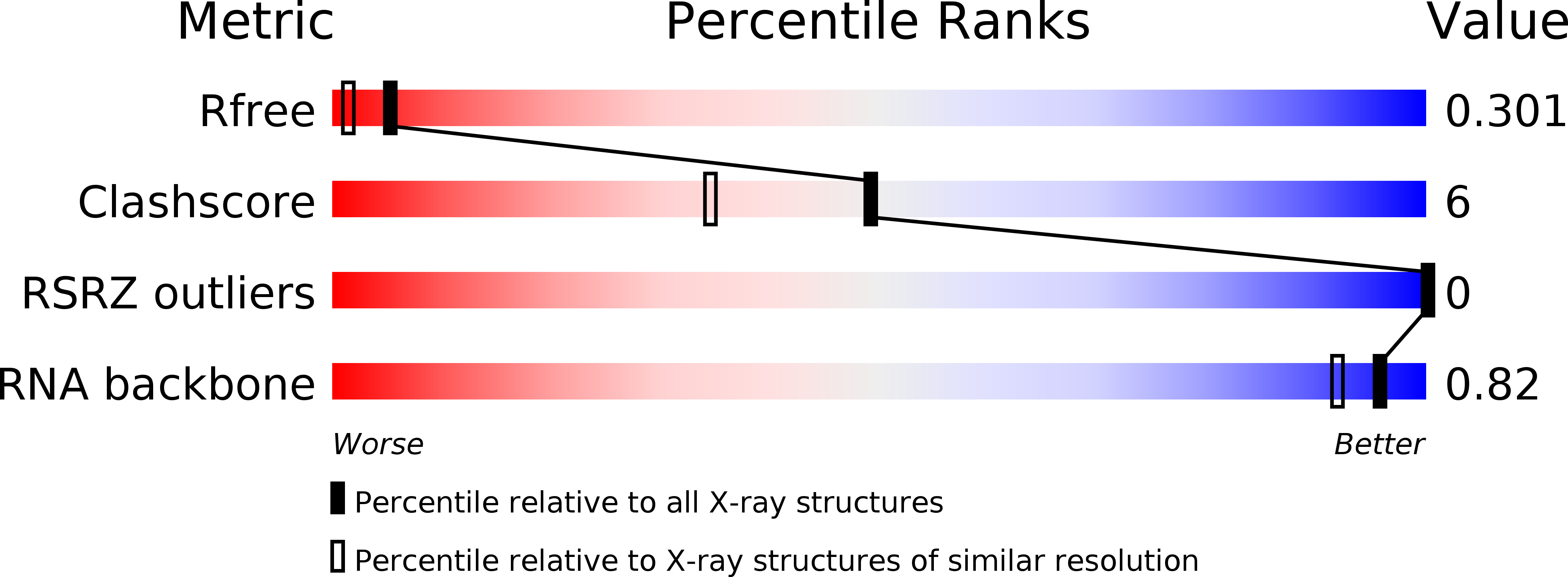
Resolution:
1.94 Å
R-Value Free:
0.29
R-Value Work:
0.22
R-Value Observed:
0.22
Space Group:
P 21 21 21
Macromolecular Entities
Polymer Type:polyribonucleotide
Molecule:RNA (5'-R(*GP*CP*AP*GP*UP*GP*GP*C)-3')
Chain IDs:A, C
Chain Length:8
Number of Molecules:2
Biological Source:
Polymer Type:polydeoxyribonucleotide/polyribonucleotide hybrid
Molecule:DNA/RNA (5'-R(*GP*CP*CP*AP)-D(*CP*TP*GP*C)-3')
Chain IDs:B, D
Chain Length:8
Number of Molecules:2
Biological Source:
Primary Citation
Crystal structure of an eight-base pair duplex containing the 3'-DNA-RNA-5' junction formed during initiation of minus-strand synthesis of HIV replication.
Biochemistry 37 12005 12011 (1998)
PMID: 9724510 DOI: 10.1021/bi981152y

Abstact

During initiation of minus-strand synthesis by HIV-1 reverse transcriptase, a 3'-DNA-RNA-5' junction is formed involving the 3'-end of tRNAlys,3. The HIV-RT-associated RNase H cleaves the RNA template strand specifically, opposite the newly synthesized DNA strand. We have determined the crystal structure at 1.9 A resolution of an eight-base pair hybrid duplex representing the junction to identify global or local structural perturbations which may be recognized by HIV-RT RNase H. The junction octamer is in a global A-type conformation throughout. A base pair step with distinct stacking geometry and variable backbone conformation is located next to the main endonucleolytic cleavage site. This base pair step may serve as a recognition site for HIV-RT RNase H.

Legend

Protein

Chemical

Disease

Primary Citation of related structures
Feedback Form
Name
Email
Institute
Feedback