Deposition Date
1997-01-03
Release Date
1997-09-22
Last Version Date
2024-04-03
Entry Detail
PDB ID:
305D
Keywords:
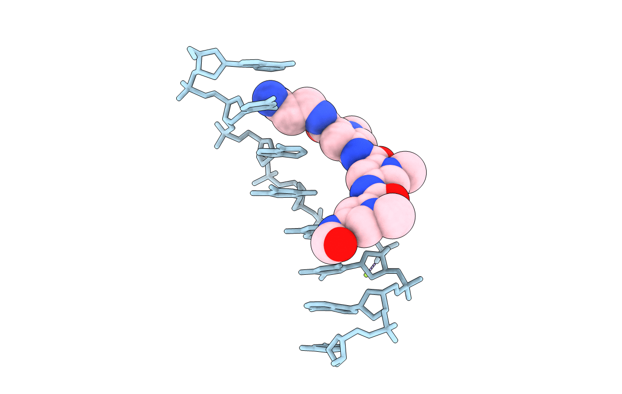
Title:
SIDE-BY-SIDE BINDING OF DISTAMYCIN MOLECULES TO D(ICATATIC) IN THE TETRAGONAL FORM
Method Details:
Experimental Method:
Resolution:
2.17 Å
R-Value Work:
0.17
R-Value Observed:
0.17
Space Group:
P 41 2 2