2zyc image
Deposition Date 2009-01-19
Release Date 2009-02-03
Last Version Date 2024-05-29
Entry Detail
PDB ID:
2ZYC
Keywords:
Title:
Crystal structure of peptidoglycan hydrolase from Sphingomonas sp. A1
Biological Source:
Source Organism:
Host Organism:
Method Details:
Experimental Method:
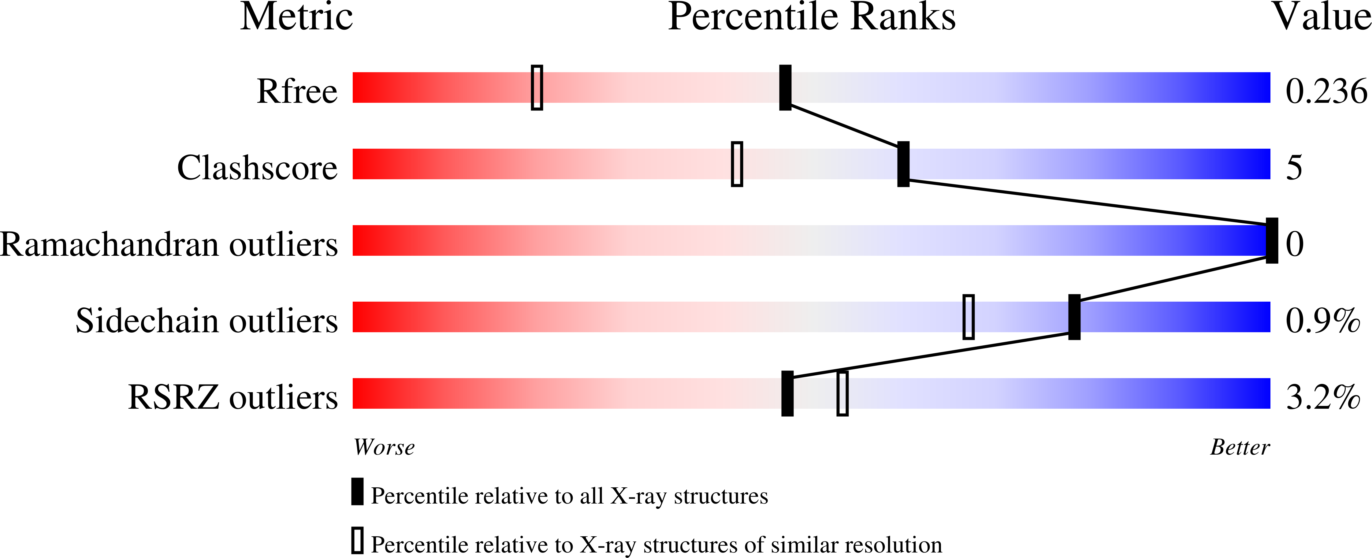
Resolution:
1.74 Å
R-Value Free:
0.23
R-Value Work:
0.19
R-Value Observed:
0.20
Space Group:
P 43 21 2
Macromolecular Entities
Structures with similar UniProt ID
Protein Blast
Polymer Type:polypeptide(L)
Molecule:Peptidoglycan hydrolase FlgJ
Gene (Uniprot):flgJ
Mutagens:T152M, P153A
Chain IDs:A
Chain Length:170
Number of Molecules:1
Biological Source:Sphingomonas sp. A1
Ligand Molecules
Primary Citation
Crystal structure of the glycosidase family 73 peptidoglycan hydrolase FlgJ
Biochem.Biophys.Res.Commun. 381 16 21 (2009)
PMID: 19351587 DOI: 10.1016/j.bbrc.2009.01.186

Abstact

Glycoside hydrolase (GH) categorized into family 73 plays an important role in degrading bacterial cell wall peptidoglycan. The flagellar protein FlgJ contains N- and C-terminal domains responsible for flagellar rod assembly and peptidoglycan hydrolysis, respectively. A member of family GH-73, the C-terminal domain (SPH1045-C) of FlgJ from Sphingomonas sp. strain A1 was expressed in Escherichia coli, purified, and characterized. SPH1045-C exhibited bacterial cell lytic activity most efficiently at pH 6.0 and 37 degrees C. The X-ray crystallographic structure of SPH1045-C was determined at 1.74 A resolution by single-wavelength anomalous diffraction. The enzyme consists of two lobes, alpha and beta. A deep cleft located between the two lobes can accommodate polymer molecules, suggesting that the active site is located in the cleft. Although SPH1045-C shows a structural homology with family GH-22 and GH-23 lysozymes, the arrangement of the nucleophile/base residue in the active site is specific to each peptidoglycan hydrolase.

Legend

Protein

Chemical

Disease

Primary Citation of related structures
Feedback Form
Name
Email
Institute
Feedback