Deposition Date
2008-12-03
Release Date
2009-12-15
Last Version Date
2023-11-01
Entry Detail
PDB ID:
2ZWD
Keywords:
Title:
Crystal structure of the copper-bound tyrosinase in complex with a caddie protein from streptomyces castaneoglobisporus obtained by soaking the deoxy-form crystal in dioxygen-saturated solution for 5 minutes
Biological Source:
Source Organism(s):
Streptomyces castaneoglobisporus (Taxon ID: 79261)
Expression System(s):
Method Details:
Experimental Method:
Resolution:
1.35 Å
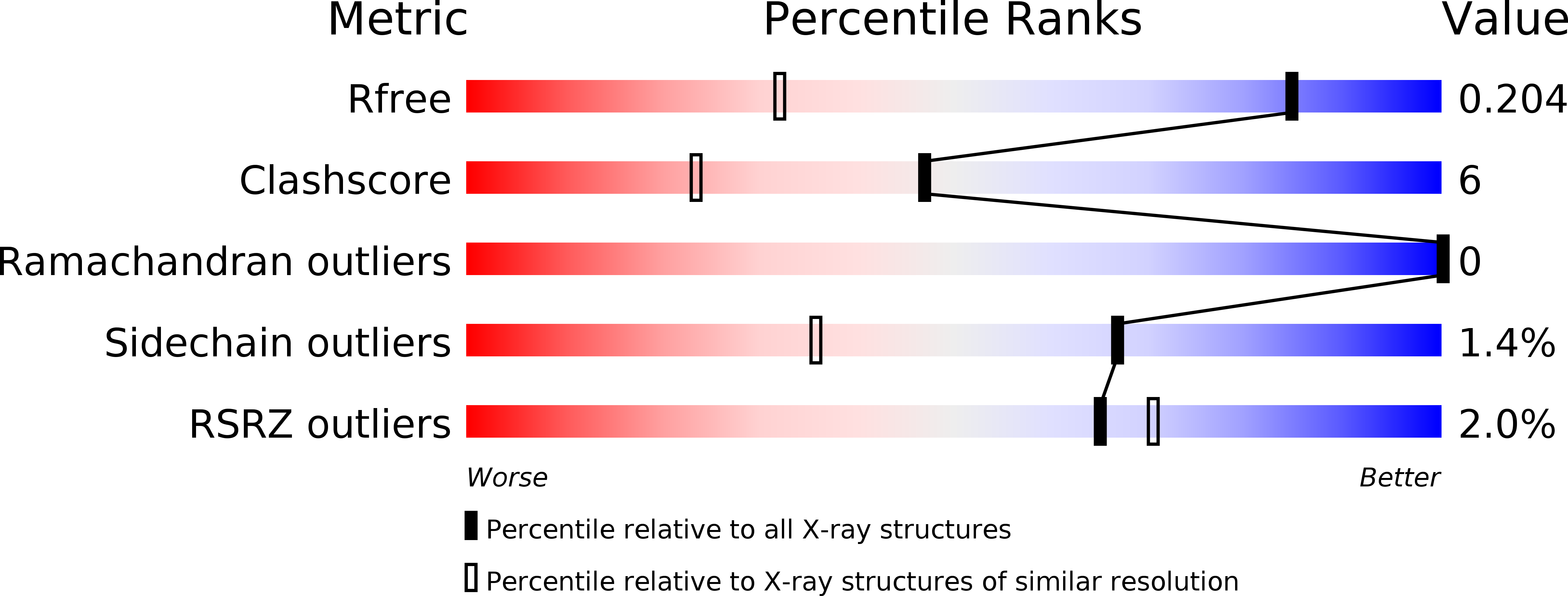
R-Value Free:
0.21
R-Value Work:
0.17
R-Value Observed:
0.17
Space Group:
P 21 21 2