Deposition Date
2008-06-09
Release Date
2009-06-09
Last Version Date
2024-11-06
Entry Detail
PDB ID:
2ZOV
Keywords:
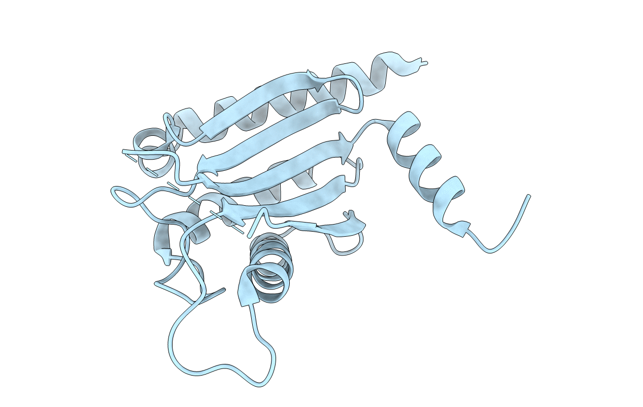
Title:
Structure of the periplasmic domain of MotB from Salmonella (crystal form I)
Biological Source:
Source Organism(s):
Salmonella typhimurium (Taxon ID: 602)
Expression System(s):
Method Details:
Experimental Method:
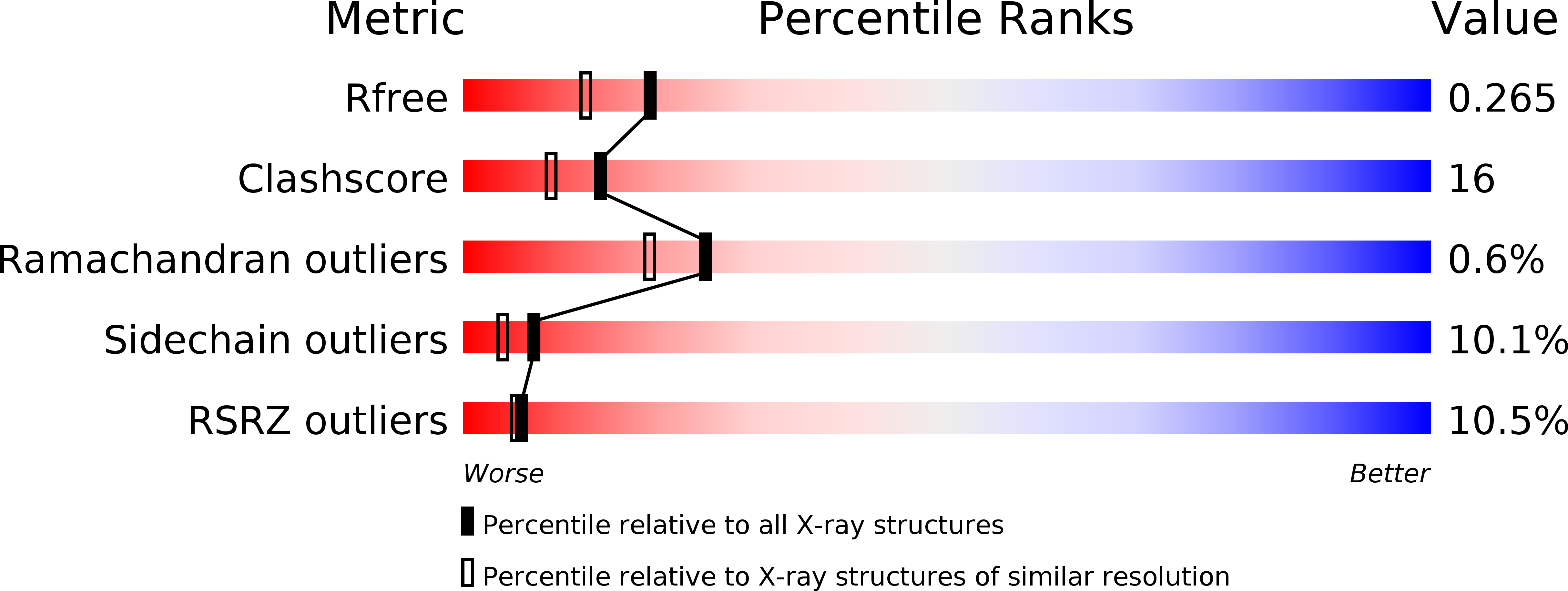
Resolution:
2.00 Å
R-Value Free:
0.26
R-Value Work:
0.23
R-Value Observed:
0.23
Space Group:
P 32 2 1