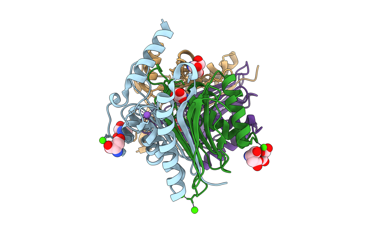
2zal image
Deposition Date 2007-10-07
Release Date 2007-10-30
Last Version Date 2023-11-01
Entry Detail
PDB ID:
2ZAL
Keywords:
Title:
Crystal structure of E. coli isoaspartyl aminopeptidase/L-asparaginase in complex with L-aspartate
Biological Source:
Source Organism:
Host Organism:
Method Details:
Experimental Method:
Resolution:
1.90 Å
R-Value Free:
0.18
R-Value Work:
0.15
R-Value Observed:
0.15
Space Group:
P 21 21 21
Macromolecular Entities
Structures with similar UniProt ID
Protein Blast
Polymer Type:polypeptide(L)
Molecule:L-asparaginase
Gene (Uniprot):iaaA
Chain IDs:A, C
Chain Length:160
Number of Molecules:2
Biological Source:Escherichia coli
Structures with similar UniProt ID
Protein Blast
Polymer Type:polypeptide(L)
Molecule:L-asparaginase
Gene (Uniprot):iaaA
Chain IDs:B, D
Chain Length:137
Number of Molecules:2
Biological Source:Escherichia coli
Primary Citation

Abstact

The crystal structure of Escherichia coli isoaspartyl aminopeptidase/asparaginase (EcAIII), an enzyme belonging to the N-terminal nucleophile (Ntn)-hydrolases family, has been determined at 1.9-A resolution for a complex obtained by cocrystallization with l-aspartate, which is a product of both enzymatic reactions catalyzed by EcAIII. The enzyme is a dimer of heterodimers, (alphabeta)(2). The (alphabeta) heterodimer, which arises by autoproteolytic cleavage of the immature protein, exhibits an alphabetabetaalpha-sandwich fold, typical for Ntn-hydrolases. The asymmetric unit contains one copy of the EcAIII.Asp complex, with clearly visible l-aspartate ligands, one bound in each of the two active sites of the enzyme. The l-aspartate ligand is located near Thr(179), the N-terminal residue of subunit beta liberated in the autoproteolytic event. Structural comparisons with the free form of EcAIII reveal that there are no major rearrangements of the active site upon aspartate binding. Although the ligand binding mode is similar to that observed in an l-aspartate complex of the related enzyme human aspartylglucosaminidase, the architecture of the EcAIII active site sheds light on the question of substrate specificity and explains why EcAIII is not able to hydrolyze glycosylated asparagine substrates.

Legend

Protein

Chemical

Disease

Primary Citation of related structures