2zak image
Deposition Date 2007-10-07
Release Date 2008-03-25
Last Version Date 2023-11-01
Entry Detail
PDB ID:
2ZAK
Keywords:
Title:
Orthorhombic crystal structure of precursor E. coli isoaspartyl peptidase/L-asparaginase (EcAIII) with active-site T179A mutation
Biological Source:
Source Organism:
Host Organism:
Method Details:
Experimental Method:
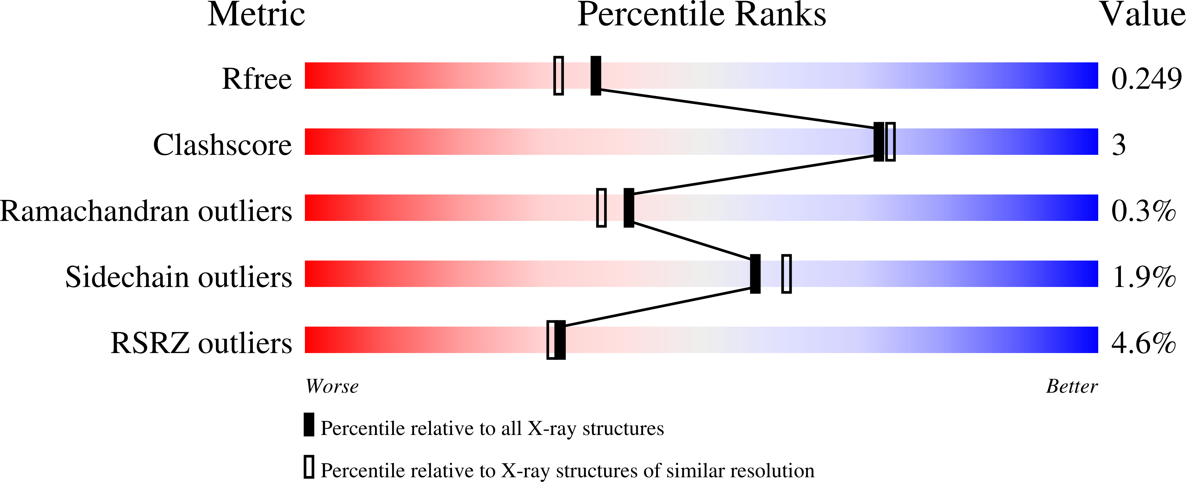
Resolution:
2.01 Å
R-Value Free:
0.25
R-Value Work:
0.19
R-Value Observed:
0.19
Space Group:
P 21 21 21
Macromolecular Entities
Structures with similar UniProt ID
Protein Blast
Polymer Type:polypeptide(L)
Molecule:L-asparaginase precursor
Gene (Uniprot):iaaA
Mutagens:T179A
Chain IDs:A, B
Chain Length:320
Number of Molecules:2
Biological Source:Escherichia coli
Primary Citation

Abstact

Plant-type L-asparaginases hydrolyze the side-chain amide bond of L-asparagine or its beta-peptides. They belong to the N-terminal nucleophile (Ntn) hydrolases and are synthesized as inactive precursor molecules. Activation occurs via the autoproteolytic release of two subunits, alpha and beta, the latter of which carries the nucleophile at its N-terminus. Crystallographic studies of plant-type asparaginases have focused on an Escherichia coli homologue (EcAIII), which has been crystallized in several crystal forms. Although they all belong to the same P2 1 2 1 2 1 space group with similar unit-cell parameters, they display different crystal-packing arrangements and thus should be classified as separate polymorphs. This variability stems mainly from different positions of the EcAIII molecules within the unit cell, although they also exhibit slight differences in orientation. The intermolecular interactions that trigger different crystal lattice formation are mediated by ions, which represent the most variable component of the crystallization conditions. This behaviour confirms recent observations that small molecules might promote protein crystal lattice formation.

Legend

Protein

Chemical

Disease

Primary Citation of related structures