Deposition Date
2007-08-31
Release Date
2007-09-11
Last Version Date
2023-12-13
Entry Detail
PDB ID:
2VAF
Keywords:
Title:
Crystal structure of Human Cardiac Calsequestrin
Biological Source:
Source Organism(s):
HOMO SAPIENS (Taxon ID: 9606)
Expression System(s):
Method Details:
Experimental Method:
Resolution:
3.80 Å
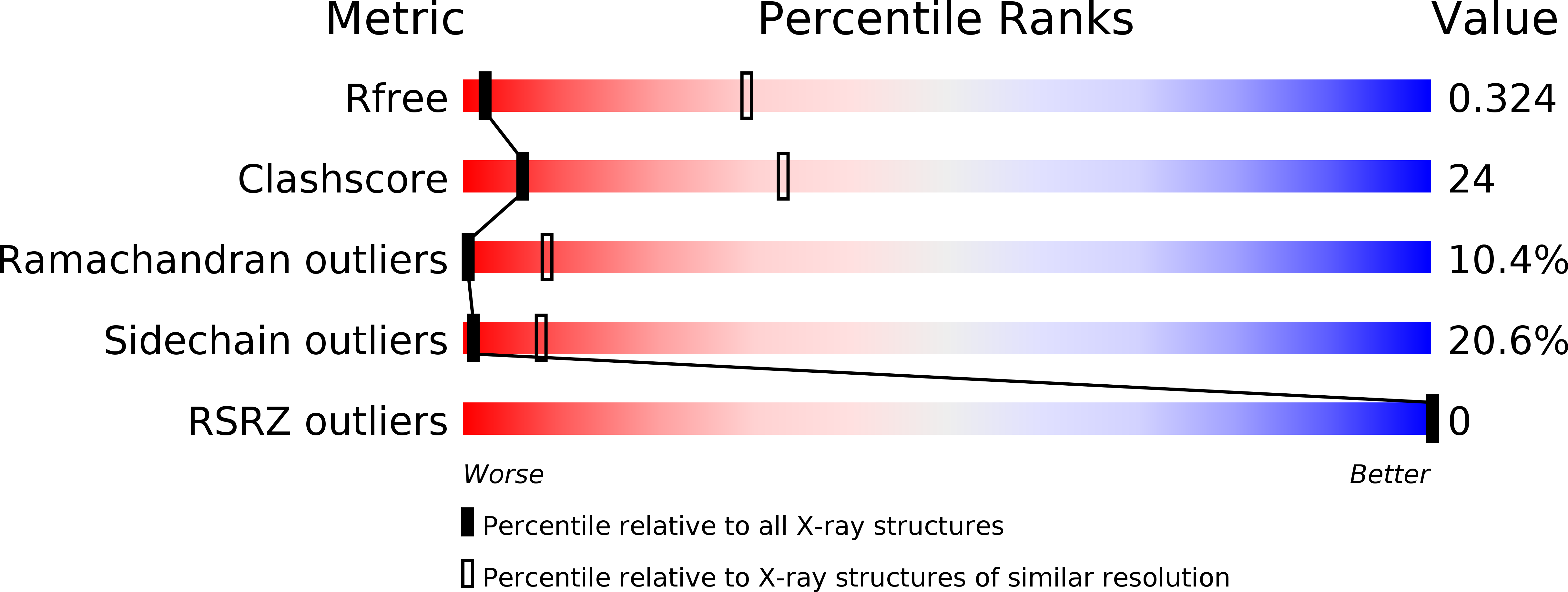
R-Value Free:
0.32
R-Value Work:
0.27
R-Value Observed:
0.27
Space Group:
I 41 2 2