2r79 image
Deposition Date 2007-09-07
Release Date 2007-10-09
Last Version Date 2024-02-21
Entry Detail
PDB ID:
2R79
Title:
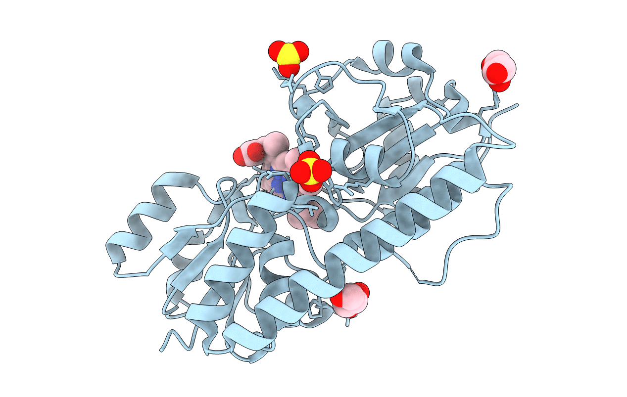
Crystal Structure of a Periplasmic Heme Binding Protein from Pseudomonas aeruginosa
Biological Source:
Source Organism:
Host Organism:
Method Details:
Experimental Method:
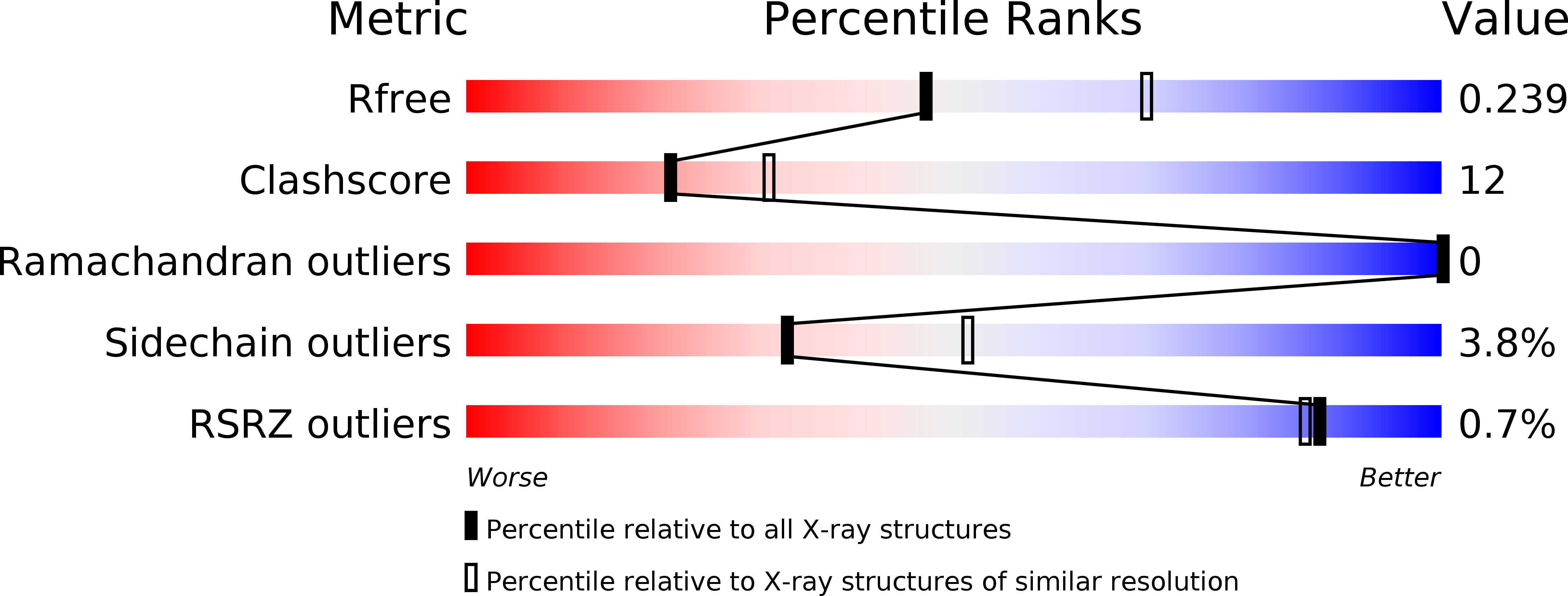
Resolution:
2.40 Å
R-Value Free:
0.24
R-Value Work:
0.19
R-Value Observed:
0.19
Space Group:
P 63 2 2
Macromolecular Entities
Polymer Type:polypeptide(L)
Molecule:Periplasmic binding protein
Gene (Uniprot):phuT
Chain IDs:A
Chain Length:283
Number of Molecules:1
Biological Source:Pseudomonas aeruginosa
Primary Citation
Holo- and apo-bound structures of bacterial periplasmic heme-binding proteins.
J.Biol.Chem. 282 35796 35802 (2007)
PMID: 17925389 DOI: 10.1074/jbc.M706761200

Abstact

An essential component of heme transport in Gram-negative bacterial pathogens is the periplasmic protein that shuttles heme between outer and inner membranes. We have solved the first crystal structures of two such proteins, ShuT from Shigella dysenteriae and PhuT from Pseudomonas aeruginosa. Both share a common architecture typical of Class III periplasmic binding proteins. The heme binds in a narrow cleft between the N- and C-terminal binding domains and is coordinated by a Tyr residue. A comparison of the heme-free (apo) and -bound (holo) structures indicates little change in structure other than minor alterations in the heme pocket and movement of the Tyr heme ligand from an "in" position where it can coordinate the heme iron to an "out" orientation where it points away from the heme pocket. The detailed architecture of the heme pocket is quite different in ShuT and PhuT. Although Arg(228) in PhuT H-bonds with a heme propionate, in ShuT a peptide loop partially takes up the space occupied by Arg(228), and there is no Lys or Arg H-bonding with the heme propionates. A comparison of PhuT/ShuT with the vitamin B(12)-binding protein BtuF and the hydroxamic-type siderophore-binding protein FhuD, the only two other structurally characterized Class III periplasmic binding proteins, demonstrates that PhuT/ShuT more closely resembles BtuF, which reflects the closer similarity in ligands, heme and B(12), compared with ligands for FhuD, a peptide siderophore.

Legend

Protein

Chemical

Disease

Primary Citation of related structures