2r0r image
Deposition Date 2007-08-21
Release Date 2008-04-29
Last Version Date 2024-11-06
Entry Detail
PDB ID:
2R0R
Title:
Crystal Structure of Human Saposin D variant SapD K9E
Biological Source:
Source Organism:
Homo sapiens (Taxon ID: 9606)
Host Organism:
Method Details:
Experimental Method:
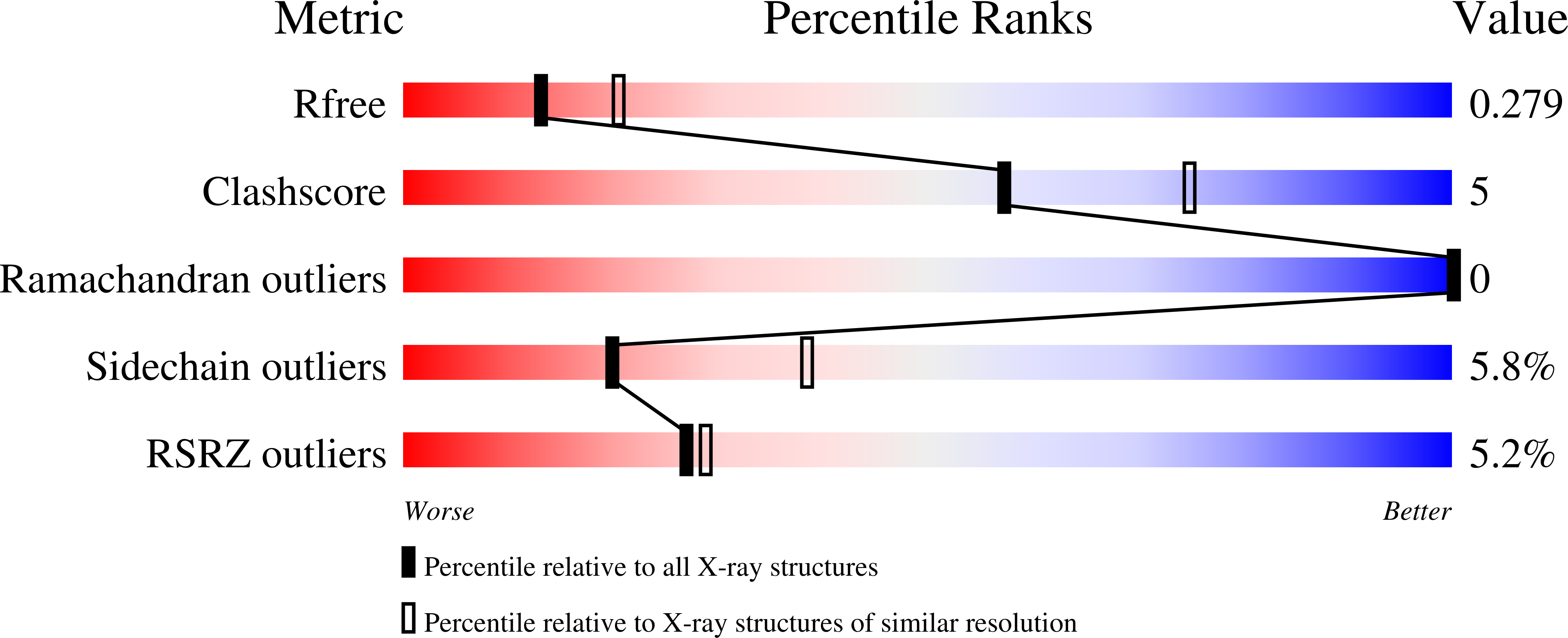
Resolution:
2.50 Å
R-Value Free:
0.28
R-Value Work:
0.21
R-Value Observed:
0.21
Space Group:
P 1 21 1
Macromolecular Entities
Structures with similar UniProt ID
Protein Blast
Polymer Type:polypeptide(L)
Molecule:Proactivator polypeptide
Gene (Uniprot):PSAP
Mutagens:K9E
Chain IDs:A, B
Chain Length:85
Number of Molecules:2
Biological Source:Homo sapiens
Ligand Molecules
Primary Citation
Crystal structures of human saposins C and d: implications for lipid recognition and membrane interactions.
Structure 16 809 817 (2008)
PMID: 18462685 DOI: 10.1016/j.str.2008.02.016

Abstact

Human saposins are essential proteins required for degradation of sphingolipids and lipid antigen presentation. Despite the conserved structural organization of saposins, their distinct modes of interaction with biological membranes are not fully understood. We describe two crystal structures of human saposin C in an "open" configuration with unusual domain swapped homodimers. This form of SapC dimer supports the "clip-on" model for SapC-induced vesicle fusion. In addition, we present the crystal structure of SapD in two crystal forms. They reveal the monomer-monomer interface for the SapD dimer, which was confirmed in solution by analytical ultracentrifugation. The crystal structure of SapD suggests that side chains of Lys10 and Arg17 are involved in initial association with the preferred anionic biological membranes by forming salt bridges with sulfate or phosphate lipid headgroups.

Legend

Protein

Chemical

Disease

Primary Citation of related structures