2qvb image
Deposition Date 2007-08-08
Release Date 2008-02-12
Last Version Date 2023-08-30
Entry Detail
PDB ID:
2QVB
Keywords:
Title:
Crystal Structure of Haloalkane Dehalogenase Rv2579 from Mycobacterium tuberculosis
Biological Source:
Source Organism:
Host Organism:
Method Details:
Experimental Method:
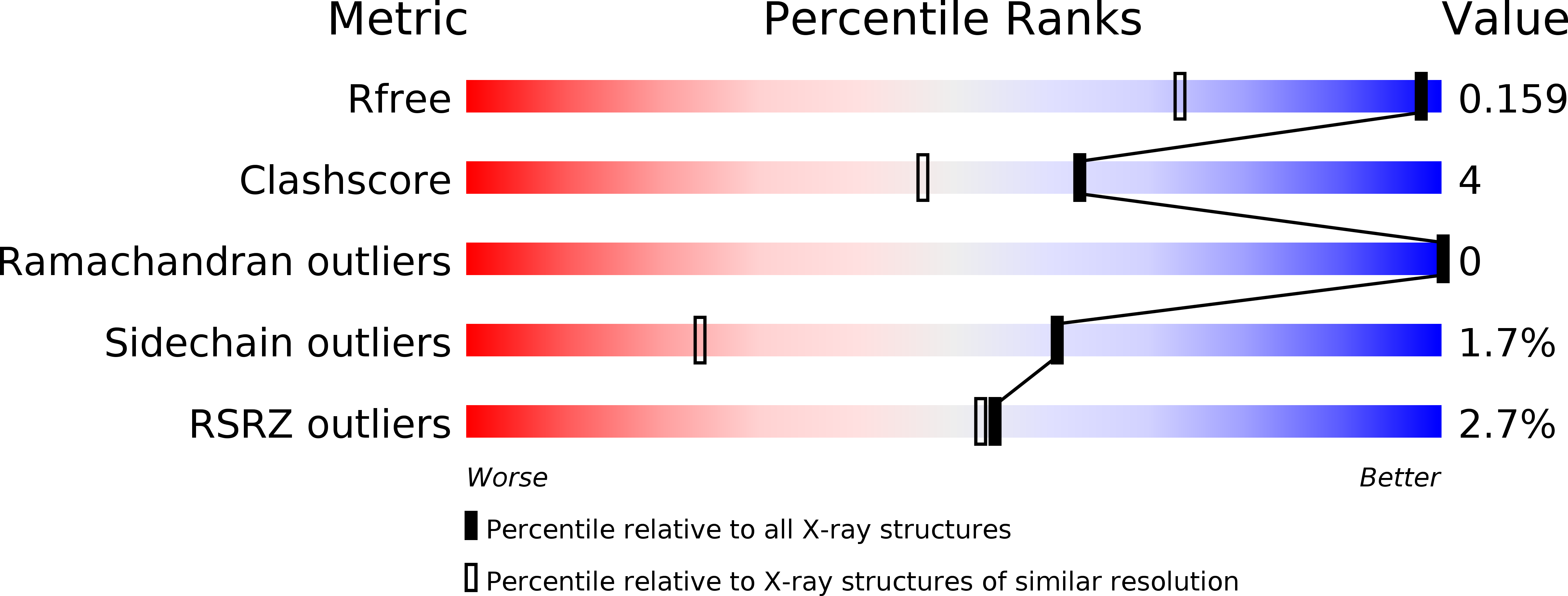
Resolution:
1.19 Å
R-Value Free:
0.16
R-Value Work:
0.12
Space Group:
P 21 21 21
Macromolecular Entities
Polymer Type:polypeptide(L)
Molecule:Haloalkane dehalogenase 3
Chain IDs:A, B
Chain Length:297
Number of Molecules:2
Biological Source:Mycobacterium tuberculosis
Primary Citation
X-ray crystal structure of Mycobacterium tuberculosis haloalkane dehalogenase Rv2579.
Biochim.Biophys.Acta 1784 351 362 (2008)
PMID: 18062934 DOI: 10.1016/j.bbapap.2007.10.014

Abstact

Haloalkane dehalogenases are enzymes well known to be important in bioremediation; the organisms from which they are produced are able to clean up toxic organohalides from polluted environments. However, besides being found in such contaminated environments, these enzymes have also been found in root or tissue-colonizing bacterial species. The haloalkane dehalogenase Rv2579 from Mycobacterium tuberculosis H37Rv has been cloned, expressed, purified and its crystal structure determined at high resolution (1.2A). In addition, the crystal structure of the enzyme has been determined in complex with the product from the reaction with 1,3-dibromopropane, i.e. 1,3-propanediol and in complex with the classical substrate of haloalkane dehalogenases, 1,2-dichloroethane. The enzyme is a two-domain protein having a catalytic domain of an alpha/beta hydrolase fold and a cap domain. The active site residues and the halide-stabilizing residues have been identified as Asp109, Glu133, His273, Asn39 and Trp110. Its overall structure is similar to those of other known haloalkane dehalogenases. Its mechanism of action involves an SN2 nucleophilic displacement.

Legend

Protein

Chemical

Disease

Primary Citation of related structures