2quo image
Deposition Date 2007-08-06
Release Date 2007-10-30
Last Version Date 2024-02-21
Entry Detail
PDB ID:
2QUO
Keywords:
Title:
Crystal Structure of C terminal fragment of Clostridium perfringens enterotoxin
Biological Source:
Source Organism:
Host Organism:
Method Details:
Experimental Method:
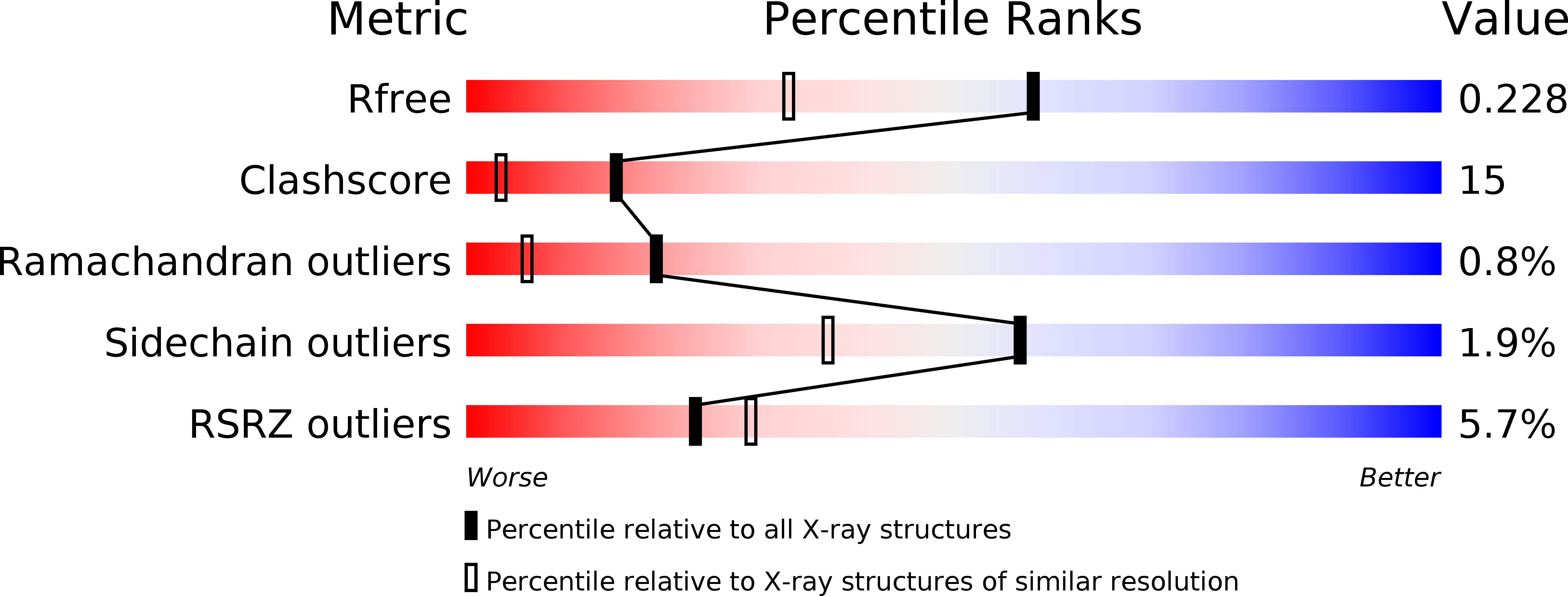
Resolution:
1.75 Å
R-Value Free:
0.22
R-Value Work:
0.19
R-Value Observed:
0.19
Space Group:
P 21 21 21
Macromolecular Entities
Structures with similar UniProt ID
Protein Blast
Polymer Type:polypeptide(L)
Molecule:Heat-labile enterotoxin B chain
Gene (Uniprot):cpe
Chain IDs:A
Chain Length:126
Number of Molecules:1
Biological Source:Clostridium perfringens
Ligand Molecules
Primary Citation
Structure of the claudin-binding domain of Clostridium perfringens enterotoxin
J.Biol.Chem. 283 268 274 (2008)
PMID: 17977833 DOI: 10.1074/jbc.M708066200

Abstact

Clostridium perfringens enterotoxin is a common cause of food-borne and antibiotic-associated diarrhea. The toxin's receptors on intestinal epithelial cells include claudin-3 and -4, members of a large family of tight junction proteins. Toxin-induced cytolytic pore formation requires residues in the NH(2)-terminal half, whereas residues near the COOH terminus are required for binding to claudins. The claudin-binding COOH-terminal domain is not toxic and is currently under investigation as a potential drug absorption enhancer. Because claudin-4 is overexpressed on some human cancers, the toxin is also being investigated for targeting chemotherapy. Our aim was to solve the structure of the claudin-binding domain to advance its therapeutic applications. The structure of a 14-kDa fragment containing residues 194 to the native COOH terminus at position 319 was solved by x-ray diffraction to a resolution of 1.75A. The structure is a nine-strand beta sandwich with previously unappreciated similarity to the receptor-binding domains of several other toxins of spore-forming bacteria, including the collagen-binding domain of ColG from Clostridium histolyticum and the large Cry family of toxins (including Cry4Ba) of Bacillus thuringiensis. Correlations with previous studies suggest that the claudin-4 binding site is on a large surface loop between strands beta8 and beta9 or includes these strands. The sequence that was crystallized (residues 194-319) binds to purified human claudin-4 with a 1:1 stoichiometry and affinity in the submicromolar range similar to that observed for binding of native toxin to cells. Our results provide a structural framework to advance therapeutic applications of the toxin and suggest a common ancestor for several receptor-binding domains of bacterial toxins.

Legend

Protein

Chemical

Disease

Primary Citation of related structures