2qq9 image
Deposition Date 2007-07-26
Release Date 2007-10-30
Last Version Date 2023-08-30
Entry Detail
PDB ID:
2QQ9
Title:
Crystal Structure of DtxR(D6A C102D) Complexed with Nickel(II)
Biological Source:
Source Organism:
Method Details:
Experimental Method:
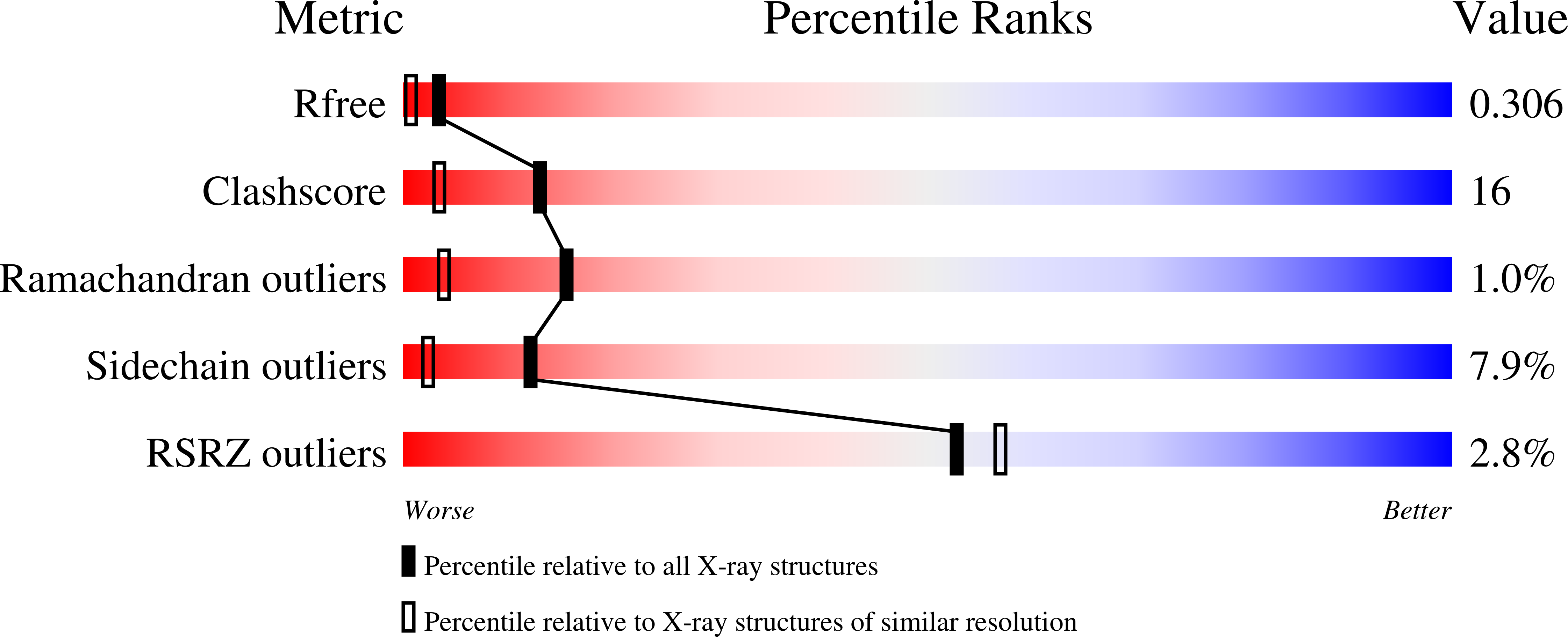
Resolution:
1.71 Å
R-Value Free:
0.29
R-Value Work:
0.23
R-Value Observed:
0.23
Space Group:
P 31 2 1
Macromolecular Entities
Structures with similar UniProt ID
Protein Blast
Polymer Type:polypeptide(L)
Molecule:Diphtheria toxin repressor
Mutagens:D6A, C102D
Chain IDs:A
Chain Length:226
Number of Molecules:1
Biological Source:Corynebacterium diphtheriae
Primary Citation
Role of the N-Terminal Helix in the Metal Ion-Induced Activation of the Diphtheria Toxin Repressor DtxR.
Biochemistry 46 11761 11770 (2007)
PMID: 17902703 DOI: 10.1021/bi7007883

Abstact

The metal ion-regulated transcriptional repressor DtxR has been shown to repress the transcription of the diphtheria toxin and other genes associated with ferrous ion homeostasis in Corynebacterium diphtheriae. In vivo studies of single-alanine mutations located in the N-terminal helix of DtxR show that the activity of the mutants is reduced compared to that of the wild type. The three-dimensional structures of the apo and activated forms of DtxR show conformational changes in the N-terminal helix resulting from metal ion activation. We have studied the N-terminal helix mutants DtxR(D6A,C102D), DtxR(E9A,C102D), and DtxR(M10A,C102D) using crystallographic and calorimetric techniques to gain insight into the possible reasons for such behavior at a molecular level. The binding affinities for metal ion extracted from the calorimetric titrations of the mutants DtxR(D6A,C102D) and DtxR(E9A,C102D) are very similar to those found for DtxR(C102D), while the same experiments performed with the mutant DtxR(M10A,C102D), bearing the M10A mutation located in binding site 2, show a decreased binding affinity in a predictable fashion. These results suggest that the decreased activity observed in these mutants cannot be explained exclusively by changes in the binding affinity of the repressor. The crystal structures of Ni-DtxR(M10A,C102D), Ni-DtxR(E9A,C102D), and Ni-DtxR(D6A,C102D) clearly show the presence of two metal ions bound. In the structure of Ni-DtxR(M10A,C102D), a water replaces Met10 in binding site 2. In the structure of Ni-DtxR(D6A,C102D), the nonhelical conformation of the N-terminal region characteristic of the activated form is absent. The side chain of Asp6 is critical in stabilization of the nonhelical conformation. This conformation is identical in all high-resolution structures of activated DtxR with an intact N-terminal helix, suggesting relevance in DtxR's regulatory function.

Legend

Protein

Chemical

Disease

Primary Citation of related structures