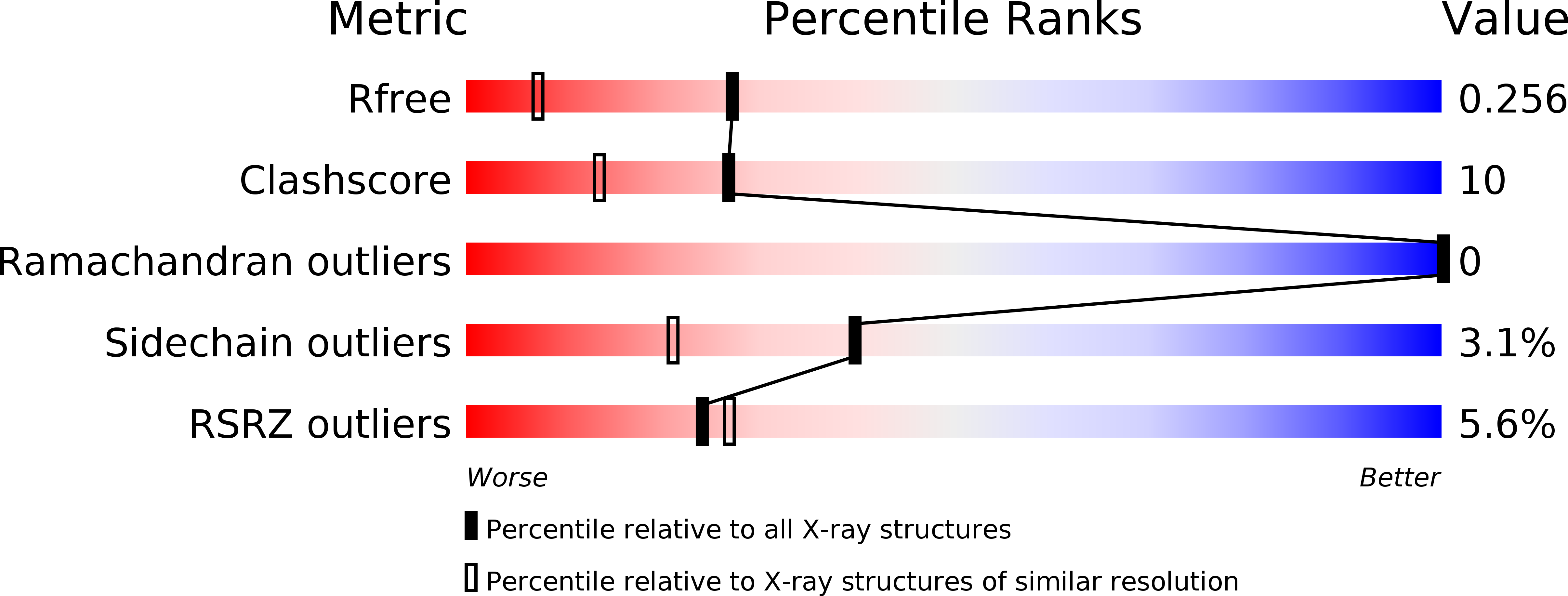
2pdr image
Deposition Date 2007-04-01
Release Date 2007-06-05
Last Version Date 2024-10-16
Entry Detail
PDB ID:
2PDR
Title:
1.7 Angstrom Crystal Structure of the Photo-excited Blue-light Photoreceptor Vivid
Biological Source:
Source Organism:
Method Details:
Experimental Method:
Resolution:
1.70 Å
R-Value Free:
0.24
R-Value Work:
0.22
R-Value Observed:
0.23
Space Group:
P 1 21 1
Macromolecular Entities
Polymer Type:polypeptide(L)
Molecule:Vivid PAS protein VVD
Gene (Uniprot):vvd
Mutations:C71S
Chain IDs:A, B, C, D
Chain Length:149
Number of Molecules:4
Biological Source:Neurospora crassa
Ligand Molecules
Primary Citation
Conformational switching in the fungal light sensor Vivid
Science 316 1054 1057 (2007)
PMID: 17510367 DOI: 10.1126/science.1137128

Abstact

The Neurospora crassa photoreceptor Vivid tunes blue-light responses and modulates gating of the circadian clock. Crystal structures of dark-state and light-state Vivid reveal a light, oxygen, or voltage Per-Arnt-Sim domain with an unusual N-terminal cap region and a loop insertion that accommodates the flavin cofactor. Photoinduced formation of a cystein-flavin adduct drives flavin protonation to induce an N-terminal conformational change. A cysteine-to-serine substitution remote from the flavin adenine dinucleotide binding site decouples conformational switching from the flavin photocycle and prevents Vivid from sending signals in Neurospora. Key elements of this activation mechanism are conserved by other photosensors such as White Collar-1, ZEITLUPE, ENVOY, and flavin-binding, kelch repeat, F-BOX 1 (FKF1).

Legend

Protein

Chemical

Disease

Primary Citation of related structures