Deposition Date
1994-06-21
Release Date
1994-12-20
Last Version Date
2023-09-27
Entry Detail
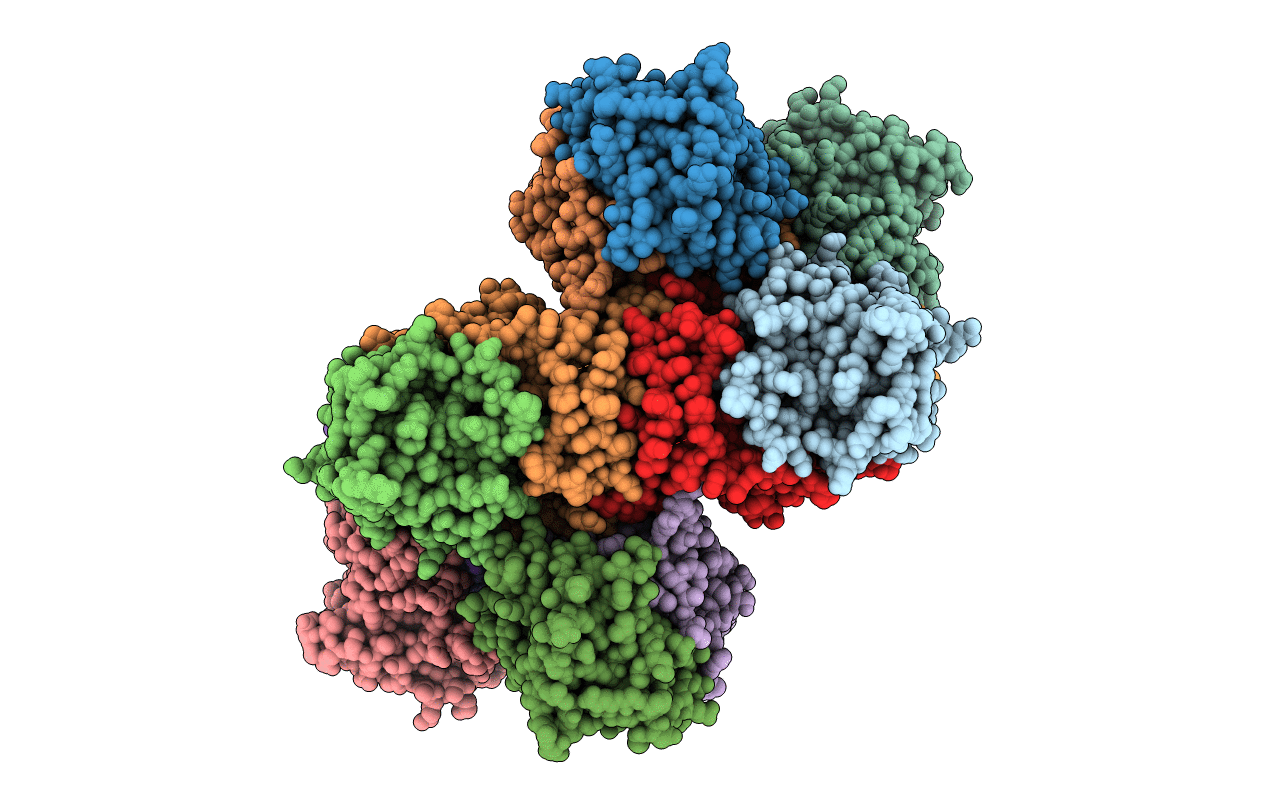
PDB ID:
2PCD
Keywords:
Title:
STRUCTURE OF PROTOCATECHUATE 3,4-DIOXYGENASE FROM PSEUDOMONAS AERUGINOSA AT 2.15 ANGSTROMS RESOLUTION
Biological Source:
Source Organism(s):
Pseudomonas putida (Taxon ID: 303)
Method Details:
Experimental Method:
Resolution:
2.15 Å
R-Value Observed:
0.17
Space Group:
I 1 2 1