2om2 image
Deposition Date 2007-01-20
Release Date 2007-07-10
Last Version Date 2023-08-30
Entry Detail
PDB ID:
2OM2
Title:
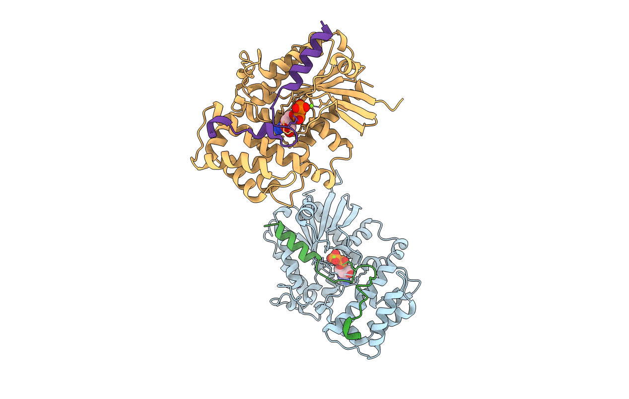
Crystal Structure Of Human G[alpha]i1 Bound To The Goloco Motif Of Rgs14
Biological Source:
Source Organism:
Homo sapiens (Taxon ID: 9606)
Host Organism:
Method Details:
Experimental Method:
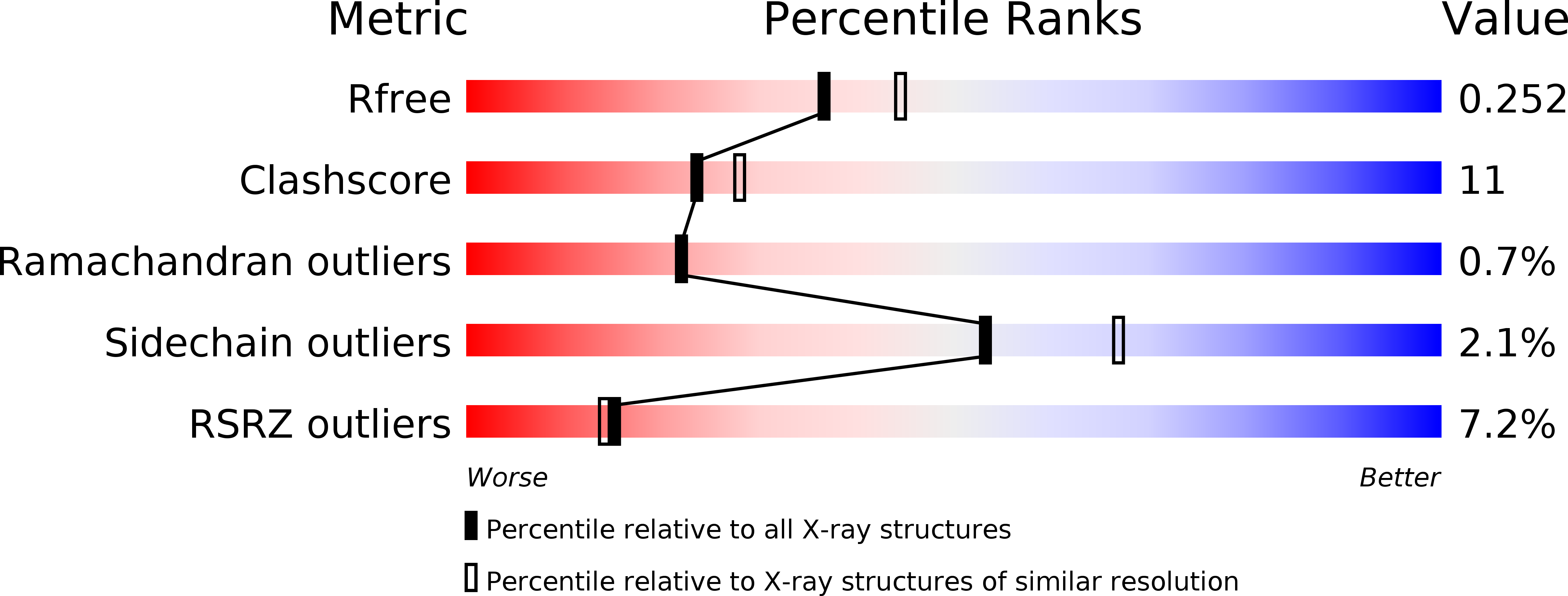
Resolution:
2.20 Å
R-Value Free:
0.26
R-Value Work:
0.22
Space Group:
P 2 2 21
Macromolecular Entities
Structures with similar UniProt ID
Protein Blast
Polymer Type:polypeptide(L)
Molecule:Guanine nucleotide-binding protein G(i), alpha-1 subunit
Gene (Uniprot):GNAI1
Chain IDs:A, C
Chain Length:325
Number of Molecules:2
Biological Source:Homo sapiens
Structures with similar UniProt ID
Protein Blast
Polymer Type:polypeptide(L)
Molecule:Regulator of G-protein signalling 14 GoLoco motif peptide
Chain IDs:B, D
Chain Length:36
Number of Molecules:2
Biological Source:
Primary Citation
Structure-based Protocol for Identifying Mutations that Enhance Protein-Protein Binding Affinities.
J.Mol.Biol. 371 1392 1404 (2007)
PMID: 17603074 DOI: 10.1016/j.jmb.2007.05.096

Abstact

The ability to manipulate protein binding affinities is important for the development of proteins as biosensors, industrial reagents, and therapeutics. We have developed a structure-based method to rationally predict single mutations at protein-protein interfaces that enhance binding affinities. The protocol is based on the premise that increasing buried hydrophobic surface area and/or reducing buried hydrophilic surface area will generally lead to enhanced affinity if large steric clashes are not introduced and buried polar groups are not left without a hydrogen bond partner. The procedure selects affinity enhancing point mutations at the protein-protein interface using three criteria: (1) the mutation must be from a polar amino acid to a non-polar amino acid or from a non-polar amino acid to a larger non-polar amino acid, (2) the free energy of binding as calculated with the Rosetta protein modeling program should be more favorable than the free energy of binding calculated for the wild-type complex and (3) the mutation should not be predicted to significantly destabilize the monomers. The performance of the computational protocol was experimentally tested on two separate protein complexes; Galpha(i1) from the heterotrimeric G-protein system bound to the RGS14 GoLoco motif, and the E2, UbcH7, bound to the E3, E6AP from the ubiquitin pathway. Twelve single-site mutations that were predicted to be stabilizing were synthesized and characterized in the laboratory. Nine of the 12 mutations successfully increased binding affinity with five of these increasing binding by over 1.0 kcal/mol. To further assess our approach we searched the literature for point mutations that pass our criteria and have experimentally determined binding affinities. Of the eight mutations identified, five were accurately predicted to increase binding affinity, further validating the method as a useful tool to increase protein-protein binding affinities.

Legend

Protein

Chemical

Disease

Primary Citation of related structures
Feedback Form
Name
Email
Institute
Feedback