2o98 image
Deposition Date 2006-12-13
Release Date 2007-04-03
Last Version Date 2023-12-27
Entry Detail
PDB ID:
2O98
Keywords:
Title:
Structure of the 14-3-3 / H+-ATPase plant complex
Biological Source:
Source Organism:
Host Organism:
Method Details:
Experimental Method:
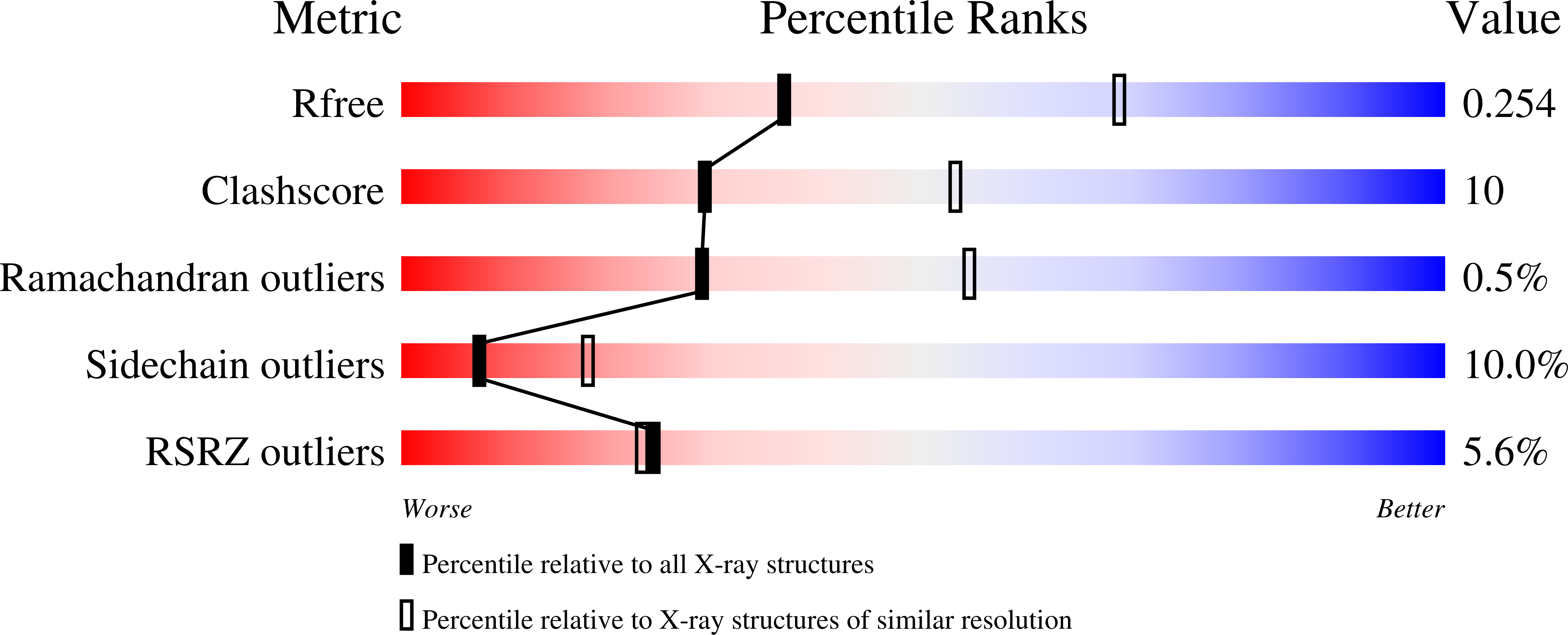
Resolution:
2.70 Å
R-Value Free:
0.26
R-Value Work:
0.17
R-Value Observed:
0.18
Space Group:
I 41 2 2
Macromolecular Entities
Structures with similar UniProt ID
Protein Blast
Polymer Type:polypeptide(L)
Molecule:14-3-3-like protein C
Chain IDs:A, B
Chain Length:242
Number of Molecules:2
Biological Source:Nicotiana tabacum
Structures with similar UniProt ID
Protein Blast
Polymer Type:polypeptide(L)
Molecule:Plasma membrane H+ ATPase
Mutagens:T439D,V440I
Chain IDs:C (auth: P), D (auth: Q)
Chain Length:52
Number of Molecules:2
Biological Source:Nicotiana plumbaginifolia
Primary Citation
Structure of a 14-3-3 coordinated hexamer of the plant plasma membrane H+ -ATPase by combining X-ray crystallography and electron cryomicroscopy
Mol.Cell 25 427 440 (2007)
PMID: 17289589 DOI: 10.1016/j.molcel.2006.12.017

Abstact

Regulatory 14-3-3 proteins activate the plant plasma membrane H(+)-ATPase by binding to its C-terminal autoinhibitory domain. This interaction requires phosphorylation of a C-terminal, mode III, recognition motif as well as an adjacent span of approximately 50 amino acids. Here we report the X-ray crystal structure of 14-3-3 in complex with the entire binding motif, revealing a previously unidentified mode of interaction. A 14-3-3 dimer simultaneously binds two H(+)-ATPase peptides, each of which forms a loop within the typical 14-3-3 binding groove and therefore exits from the center of the dimer. Several H(+)-ATPase mutants support this structure determination. Accordingly, 14-3-3 binding could result in H(+)-ATPase oligomerization. Indeed, by using single-particle electron cryomicroscopy, the 3D reconstruction of the purified H(+)-ATPase/14-3-3 complex demonstrates a hexameric arrangement. Fitting of 14-3-3 and H(+)-ATPase atomic structures into the 3D reconstruction map suggests the spatial arrangement of the holocomplex.

Legend

Protein

Chemical

Disease

Primary Citation of related structures