Deposition Date
2008-03-07
Release Date
2008-09-09
Last Version Date
2024-05-29
Entry Detail
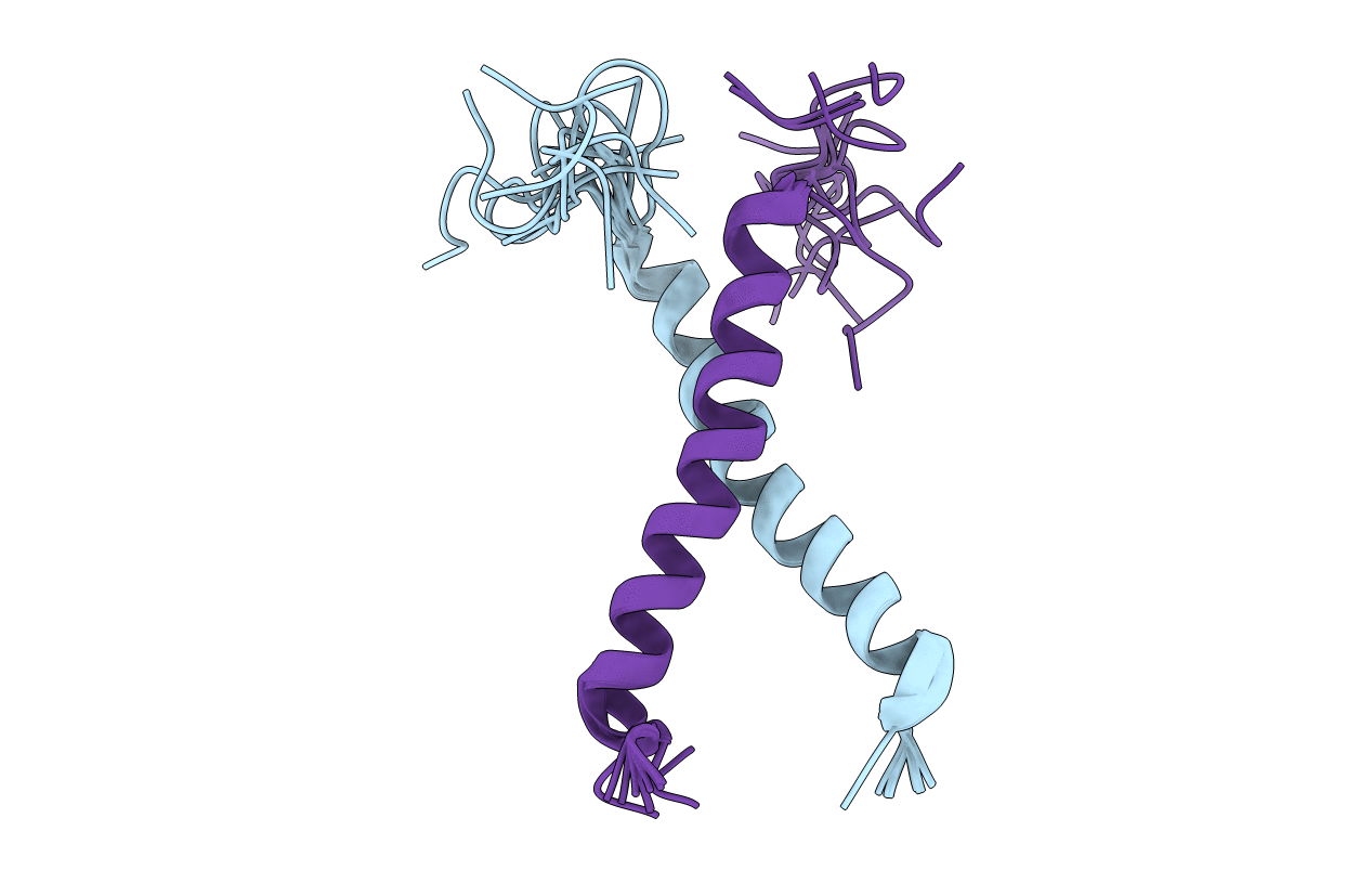
PDB ID:
2K1L
Keywords:
Title:
NMR structures of dimeric transmembrane domain of the receptor tyrosine kinase EphA1 in lipid bicelles at pH 6.3
Biological Source:
Source Organism(s):
Homo sapiens (Taxon ID: 9606)
Expression System(s):
Method Details:
Experimental Method:
Conformers Calculated:
100
Conformers Submitted:
12
Selection Criteria:
target function