Deposition Date
2008-02-13
Release Date
2008-11-11
Last Version Date
2024-05-29
Entry Detail
PDB ID:
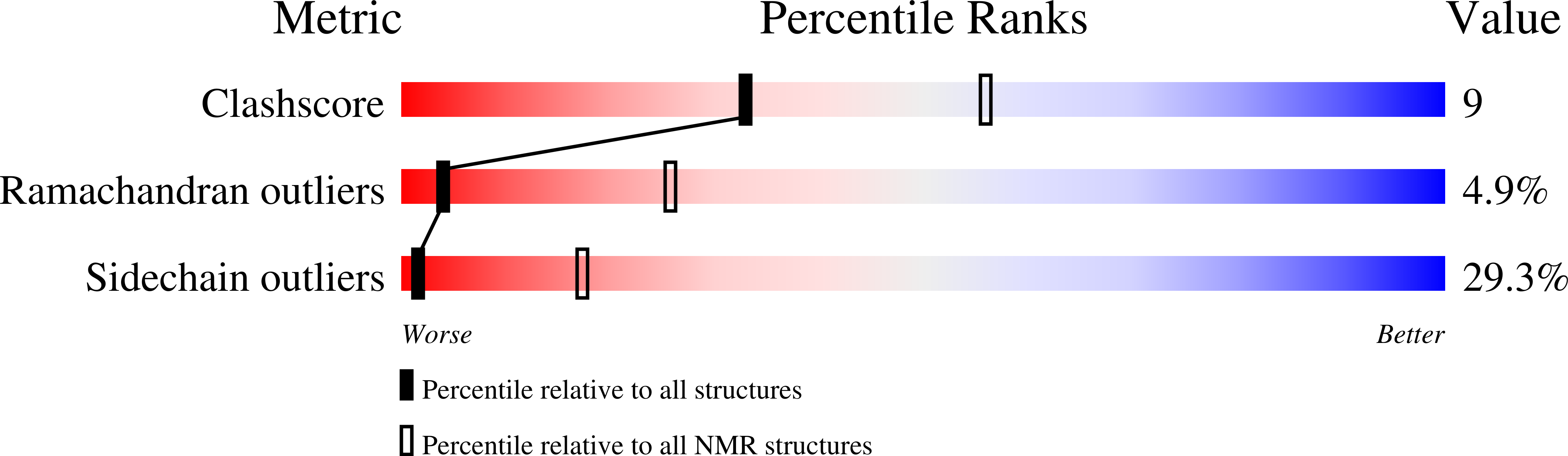
2K0R
Keywords:
Title:
Solution structure of the C103S mutant of the N-terminal Domain of DsbD from Neisseria meningitidis
Biological Source:
Source Organism(s):
Neisseria meningitidis serogroup B (Taxon ID: 491)
Expression System(s):
Method Details:
Experimental Method:
Conformers Calculated:
1500
Conformers Submitted:
20
Selection Criteria:
target function