Deposition Date
2008-01-04
Release Date
2008-02-19
Last Version Date
2024-05-08
Entry Detail
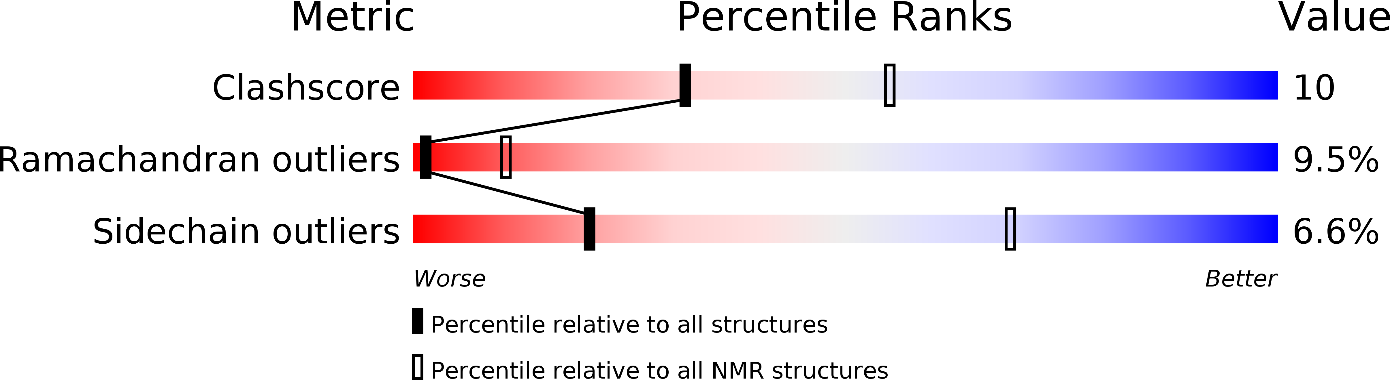
PDB ID:
2JZC
Keywords:
Title:
NMR solution structure of ALG13: The sugar donor subunit of a yeast N-acetylglucosamine transferase. Northeast Structural Genomics Consortium target YG1
Biological Source:
Source Organism(s):
Saccharomyces cerevisiae (Taxon ID: 4932)
Expression System(s):
Method Details:
Experimental Method:
Conformers Calculated:
50
Conformers Submitted:
10
Selection Criteria:
structures with the least restraint violations