Deposition Date
2006-10-13
Release Date
2007-08-28
Last Version Date
2024-11-13
Entry Detail
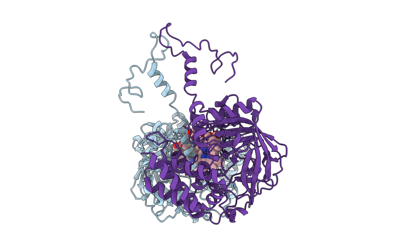
PDB ID:
2IQF
Keywords:
Title:
Crystal structure of Helicobacter pylori catalase compound I
Biological Source:
Source Organism(s):
Helicobacter pylori (Taxon ID: 210)
Expression System(s):
Method Details:
Experimental Method:
Resolution:
1.86 Å
R-Value Free:
0.18
R-Value Work:
0.14
R-Value Observed:
0.15
Space Group:
P 21 21 2