2img image
Deposition Date 2006-10-04
Release Date 2006-10-17
Last Version Date 2024-10-30
Entry Detail
PDB ID:
2IMG
Keywords:
Title:
Crystal structure of dual specificity protein phosphatase 23 from Homo sapiens in complex with ligand malate ion
Biological Source:
Source Organism(s):
Homo sapiens (Taxon ID: 9606)
Expression System(s):
Method Details:
Experimental Method:
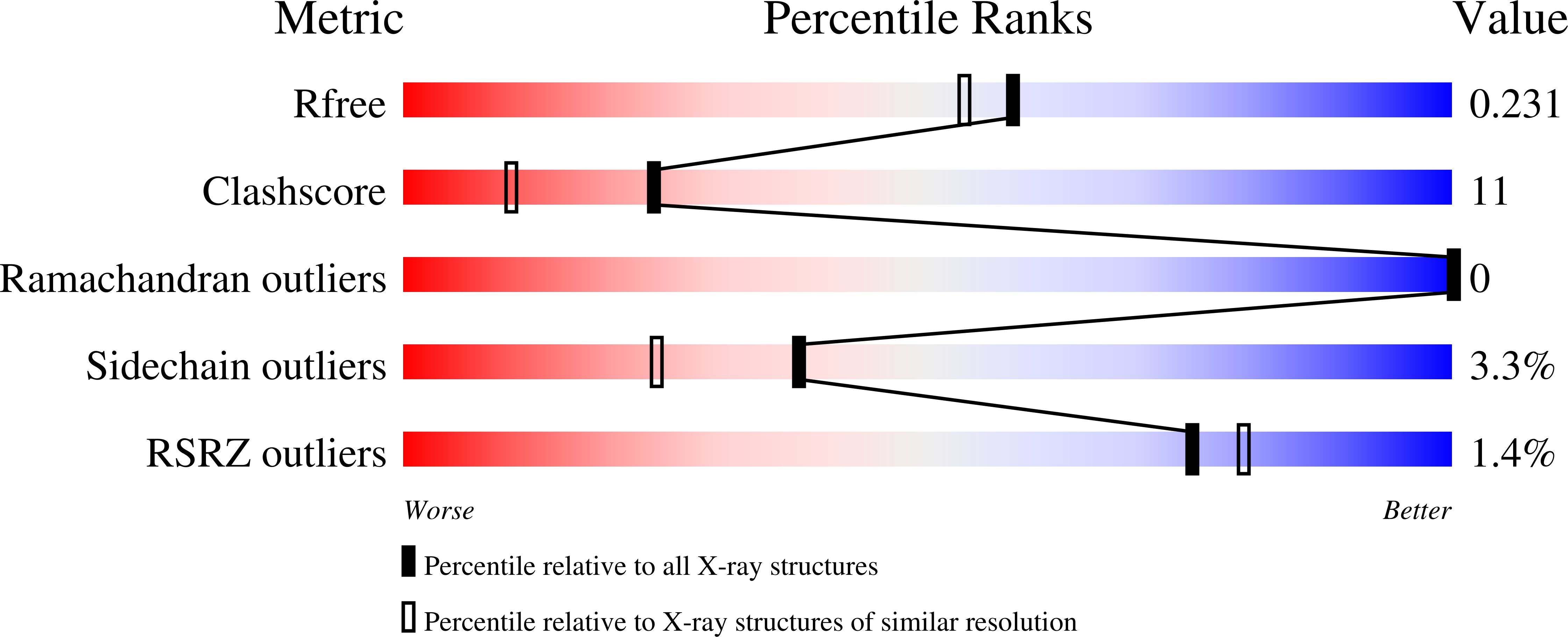
Resolution:
1.93 Å
R-Value Free:
0.22
R-Value Work:
0.19
R-Value Observed:
0.19
Space Group:
P 21 21 21
Macromolecular Entities
Structures with similar UniProt ID
Protein Blast
Polymer Type:polypeptide(L)
Molecule:Dual specificity protein phosphatase 23
Gene (Uniprot):DUSP23
Chain IDs:A
Chain Length:151
Number of Molecules:1
Biological Source:Homo sapiens
Modified Residue
Compound ID Chain ID Parent Comp ID Details 2D Image
MSE A MET SELENOMETHIONINE
Ligand Molecules
Primary Citation
Structure of human dual specificity protein phosphatase 23, VHZ, enzyme-substrate/product complex.
J.Biol.Chem. 283 8946 8953 (2008)
PMID: 18245086 DOI: 10.1074/jbc.M708945200

Abstact

Protein phosphorylation plays a crucial role in mitogenic signal transduction and regulation of cell growth and differentiation. Dual specificity protein phosphatase 23 (DUSP23) or VHZ mediates dephosphorylation of phospho-tyrosyl (pTyr) and phospho-seryl/threonyl (pSer/pThr) residues in specific proteins. In vitro, it can dephosphorylate p44ERK1 but not p54SAPK-beta and enhance activation of c-Jun N-terminal kinase (JNK) and p38. Human VHZ, the smallest of the catalytically active protein-tyrosine phosphatases (PTP) reported to date (150 residues), is a class I Cys-based PTP and bears the distinctive active site signature motif HCXXGXXRS(T). We present the crystal structure of VHZ determined at 1.93A resolution. The polypeptide chain adopts the typical alphabetaalpha PTP fold, giving rise to a shallow active site cleft that supports dual phosphorylated substrate specificity. Within our crystals, the Thr-135-Tyr-136 from a symmetry-related molecule bind in the active site with a malate ion, where they mimic the phosphorylated TY motif of the MAPK activation loop in an enzyme-substrate/product complex. Analyses of intermolecular interactions between the enzyme and this pseudo substrate/product along with functional analysis of Phe-66, Leu-97, and Phe-99 residues provide insights into the mechanism of substrate binding and catalysis in VHZ.

Legend

Protein

Chemical

Disease

Primary Citation of related structures
Feedback Form
Name
Email
Institute
Feedback