
Deposition Date
1994-09-19
Release Date
1994-12-20
Last Version Date
2024-02-14
Entry Detail
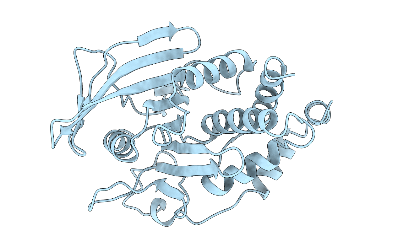
PDB ID:
2HNP
Keywords:
Title:
CRYSTAL STRUCTURE OF HUMAN PROTEIN TYROSINE PHOSPHATASE 1B
Biological Source:
Source Organism(s):
Homo sapiens (Taxon ID: 9606)
Method Details:
Experimental Method:
Resolution:
2.85 Å
R-Value Work:
0.20
R-Value Observed:
0.20
Space Group:
P 31 2 1
