2hig image
Deposition Date 2006-06-29
Release Date 2007-02-13
Last Version Date 2024-02-14
Entry Detail
PDB ID:
2HIG
Keywords:
Title:
Crystal Structure of Phosphofructokinase apoenzyme from Trypanosoma brucei.
Biological Source:
Source Organism:
Host Organism:
Method Details:
Experimental Method:
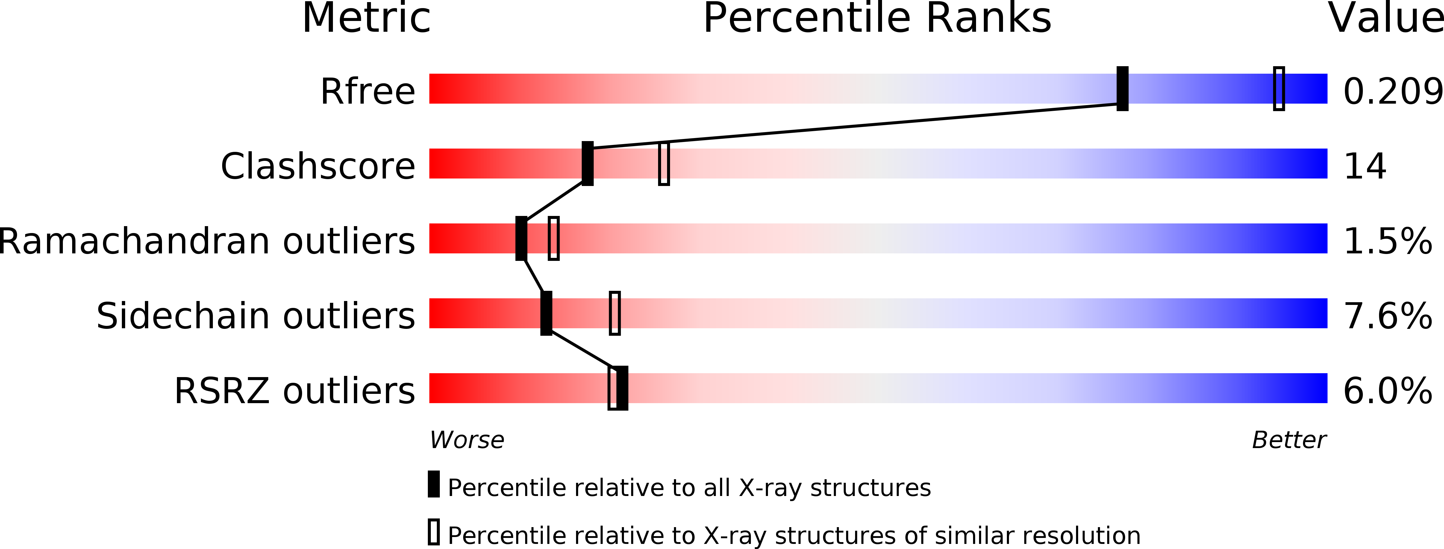
Resolution:
2.40 Å
R-Value Free:
0.23
R-Value Work:
0.20
R-Value Observed:
0.20
Space Group:
P 21 21 2
Macromolecular Entities
Structures with similar UniProt ID
Protein Blast
Polymer Type:polypeptide(L)
Molecule:6-phospho-1-fructokinase
Gene (Uniprot):pfk
Chain IDs:A, B
Chain Length:487
Number of Molecules:2
Biological Source:Trypanosoma brucei
Ligand Molecules
Primary Citation
The First Crystal Structure of Phosphofructokinase from a Eukaryote: Trypanosoma brucei.
J.Mol.Biol. 366 1185 1198 (2007)
PMID: 17207816 DOI: 10.1016/j.jmb.2006.10.019

Abstact

The crystal structure of the ATP-dependent phosphofructokinase (PFK) from Trypanosoma brucei provides the first detailed description of a eukaryotic PFK, and enables comparisons to be made with the crystal structures of bacterial ATP-dependent and PPi-dependent PFKs. The structure reveals that two insertions (the 17-20 and 329-348 loops) that are characteristic of trypanosomatid PFKs, but absent from bacterial and mammalian ATP-dependent PFKs, are located within and adjacent to the active site, and are in positions to play important roles in the enzyme's mechanism. The 90 residue N-terminal extension forms a novel domain that includes an "embracing arm" across the subunit boundary to the symmetry-related subunit in the tetrameric enzyme. Comparisons with the PPi-dependent PFK from Borrelia burgdorferi show that several features thought to be characteristic of PPi-dependent PFKs are present in the trypanosome ATP-dependent PFK. These two enzymes are generally more similar to each other than to the bacterial or mammalian ATP-dependent PFKs. However, there are critical differences at the active site of PPi-dependent PFKs that are sufficient to prevent the binding of ATP. This crystal structure of a eukaryotic PFK has enabled us to propose a detailed model of human muscle PFK that shows active site and other differences that offer opportunities for structure-based drug discovery for the treatment of sleeping sickness and other diseases caused by the trypanosomatid family of protozoan parasites.

Legend

Protein

Chemical

Disease

Primary Citation of related structures
Feedback Form
Name
Email
Institute
Feedback