2gq9 image
Deposition Date 2006-04-20
Release Date 2006-07-25
Last Version Date 2024-02-14
Entry Detail
PDB ID:
2GQ9
Keywords:
Title:
Structure of SYE1, an OYE homologue from S. oneidensis, in complex with p-hydroxybenzaldehyde
Biological Source:
Source Organism:
Method Details:
Experimental Method:
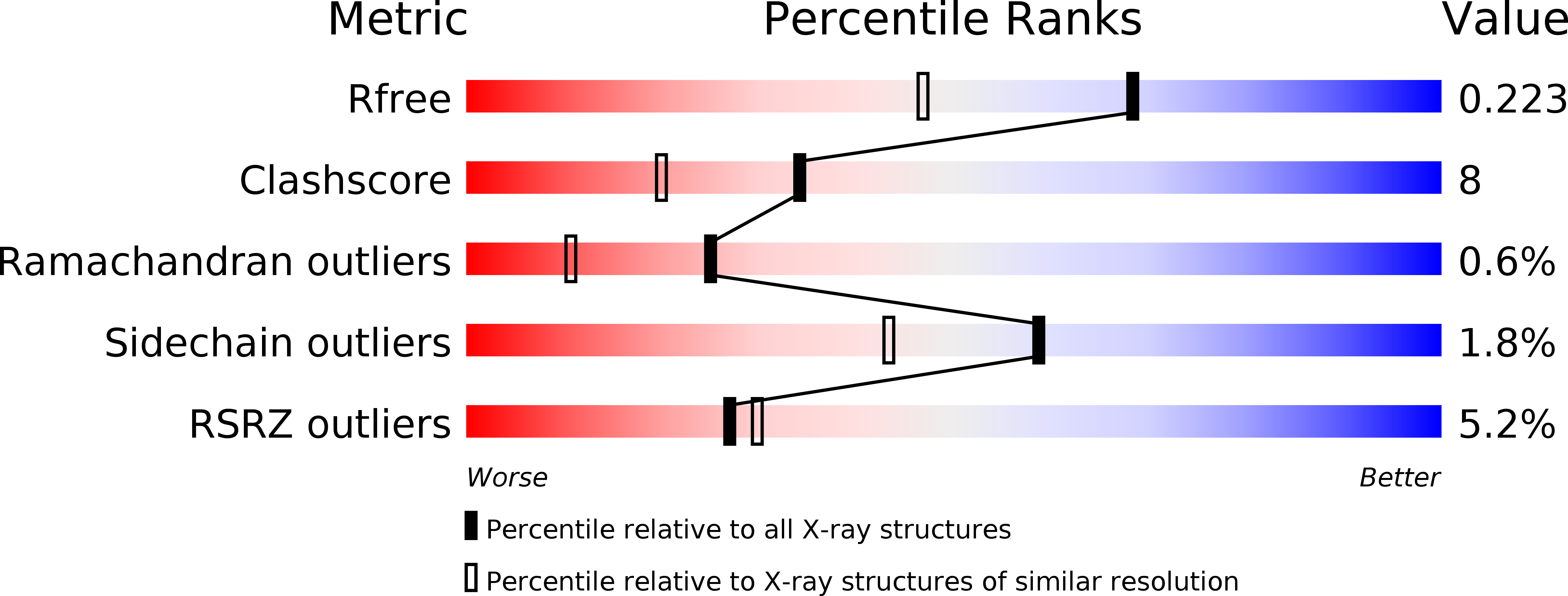
Resolution:
1.70 Å
R-Value Free:
0.22
R-Value Work:
0.18
R-Value Observed:
0.19
Space Group:
P 21 21 21
Macromolecular Entities
Structures with similar UniProt ID
Protein Blast
Polymer Type:polypeptide(L)
Molecule:oxidoreductase, FMN-binding
Gene (Uniprot):sye1
Chain IDs:A
Chain Length:365
Number of Molecules:1
Biological Source:Shewanella oneidensis
Primary Citation
Ligand-induced conformational changes in the capping subdomain of a bacterial old yellow enzyme homologue and conserved sequence fingerprints provide new insights into substrate binding.
J.Biol.Chem. 281 28152 28161 (2006)
PMID: 16857682 DOI: 10.1074/jbc.M603946200

Abstact

We have recently reported that Shewanella oneidensis, a Gram-negative gamma-proteobacterium with a rich arsenal of redox proteins, possesses four old yellow enzyme (OYE) homologues. Here, we report a series of high resolution crystal structures for one of these OYEs, Shewanella yellow enzyme 1 (SYE1), in its oxidized form at 1.4A resolution, which binds a molecule of PEG 400 in the active site, and in its NADH-reduced and p-hydroxybenzaldehyde- and p-hydroxyacetophenone-bound forms at 1.7A resolution. Although the overall structure of SYE1 reveals a monomeric enzyme based on the alpha(8)beta(8) barrel scaffold observed for other OYEs, the active site exhibits a unique combination of features: a strongly butterfly-bent FMN cofactor both in the oxidized and NADH-reduced forms, a collapsed and narrow active site tunnel, and a novel combination of conserved residues involved in the binding of phenolic ligands. Furthermore, we identify a second p-hydroxybenzaldehyde-binding site in a hydrophobic cleft next to the entry of the active site tunnel in the capping subdomain, formed by a restructuring of Loop 3 to an "open" conformation. This constitutes the first evidence to date for the entire family of OYEs that Loop 3 may indeed play a dynamic role in ligand binding and thus provides insights into the elusive NADH complex and into substrate binding in general. Structure-based sequence alignments indicate that the novelties we observe in SYE1 are supported by conserved residues in a number of structurally uncharacterized OYEs from the beta- and gamma-proteobacteria, suggesting that SYE1 represents a new subfamily of bacterial OYEs.

Legend

Protein

Chemical

Disease

Primary Citation of related structures
Feedback Form
Name
Email
Institute
Feedback