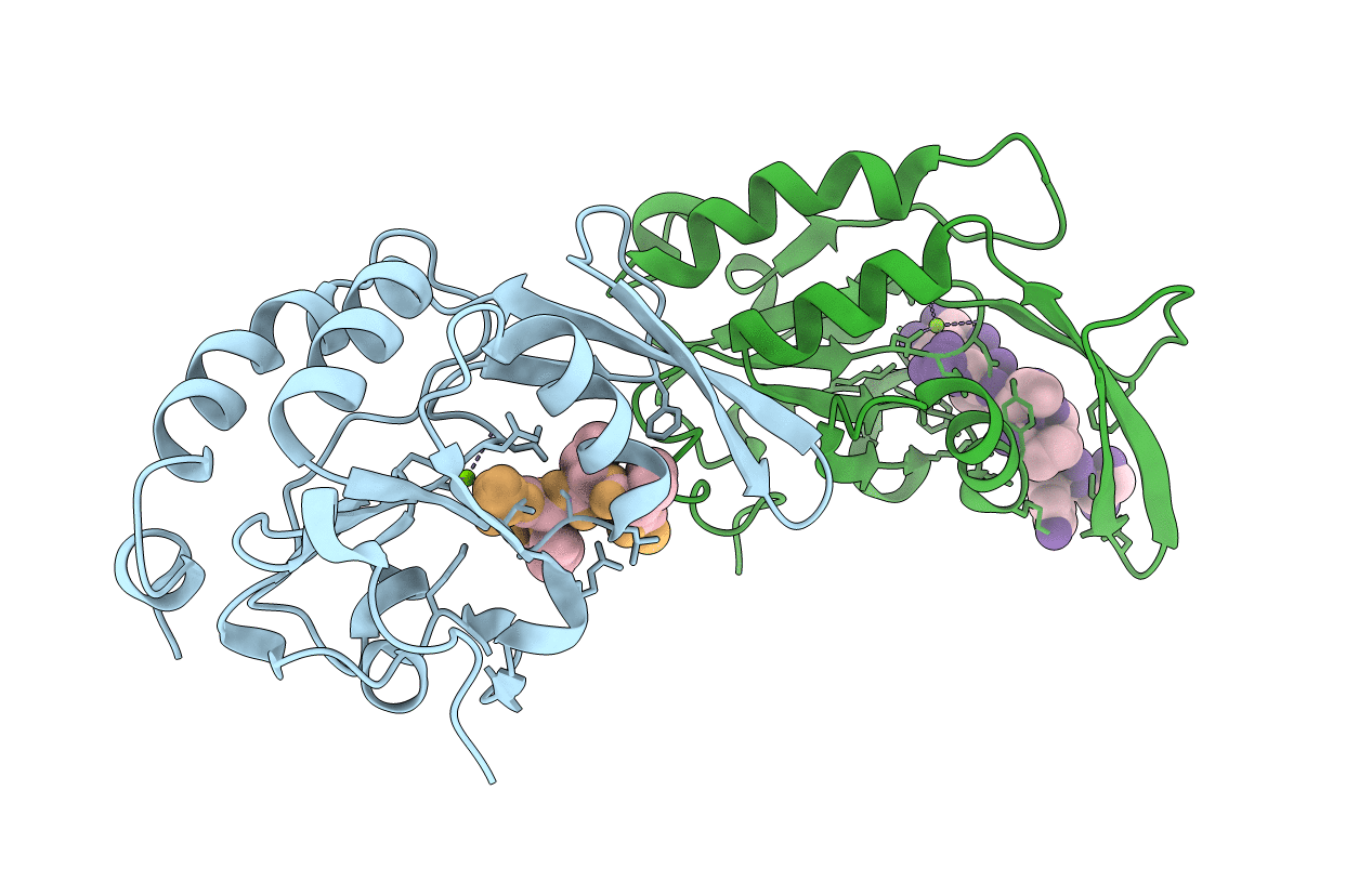
2ght image
Deposition Date 2006-03-27
Release Date 2006-12-05
Last Version Date 2024-10-30
Entry Detail
PDB ID:
2GHT
Keywords:
Title:
CTD-specific phosphatase Scp1 in complex with peptide from C-terminal domain of RNA polymerase II
Biological Source:
Source Organism:
Homo sapiens (Taxon ID: 9606)
Method Details:
Experimental Method:
Resolution:
1.80 Å
R-Value Free:
0.23
R-Value Work:
0.21
R-Value Observed:
0.24
Space Group:
C 1 2 1
Macromolecular Entities
Structures with similar UniProt ID
Protein Blast
Polymer Type:polypeptide(L)
Molecule:Carboxy-terminal domain RNA polymerase II polypeptide A small phosphatase 1
Gene (Uniprot):CTDSP1
Mutagens:D96N
Chain IDs:A, B
Chain Length:181
Number of Molecules:2
Biological Source:Homo sapiens
Structures with similar UniProt ID
Protein Blast
Polymer Type:polypeptide(L)
Molecule:DNA-directed RNA polymerase II largest subunit
Gene (Uniprot):POLR2A
Chain IDs:C, D
Chain Length:8
Number of Molecules:2
Biological Source:Homo sapiens
Modified Residue
Compound ID Chain ID Parent Comp ID Details 2D Image
SEP C SER PHOSPHOSERINE
Ligand Molecules
Primary Citation
Determinants for dephosphorylation of the RNA polymerase II C-terminal domain by Scp1.
Mol.Cell 24 759 770 (2006)
PMID: 17157258 DOI: 10.1016/j.molcel.2006.10.027

Abstact

Phosphorylation and dephosphorylation of the C-terminal domain (CTD) of RNA polymerase II (Pol II) represent a critical regulatory checkpoint for transcription. Transcription initiation requires Fcp1/Scp1-mediated dephosphorylation of phospho-CTD. Fcp1 and Scp1 belong to a family of Mg2+ -dependent phosphoserine (P.Ser)/phosphothreonine (P.Thr)-specific phosphatases. We recently showed that Scp1 is an evolutionarily conserved regulator of neuronal gene silencing. Here, we present the X-ray crystal structures of a dominant-negative form of human Scp1 (D96N mutant) bound to mono- and diphosphorylated peptides encompassing the CTD heptad repeat (Y1S2P3T4S5P6S7). Moreover, kinetic and thermodynamic analyses of Scp1-phospho-CTD peptide complexes support the structures determined. This combined structure-function analysis discloses the residues in Scp1 involved in CTD binding and its preferential dephosphorylation of P.Ser5 of the CTD heptad repeat. Moreover, these results provide a template for the design of specific inhibitors of Scp1 for the study of neuronal stem cell development.

Legend

Protein

Chemical

Disease

Primary Citation of related structures