2g2p image
Deposition Date 2006-02-16
Release Date 2006-12-12
Last Version Date 2023-10-25
Entry Detail
PDB ID:
2G2P
Title:
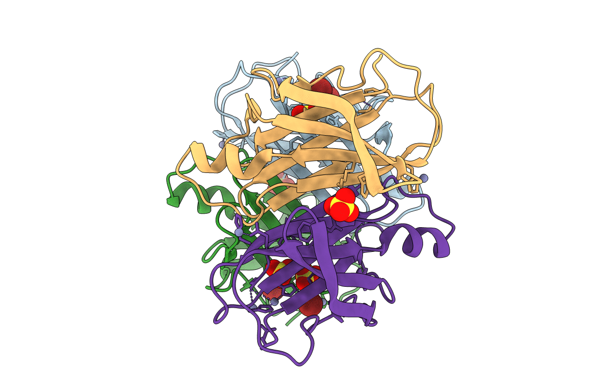
Crystal Structure of E.coli transthyretin-related protein with bound Zn and Br
Biological Source:
Source Organism:
Escherichia coli (Taxon ID: 562)
Host Organism:
Method Details:
Experimental Method:
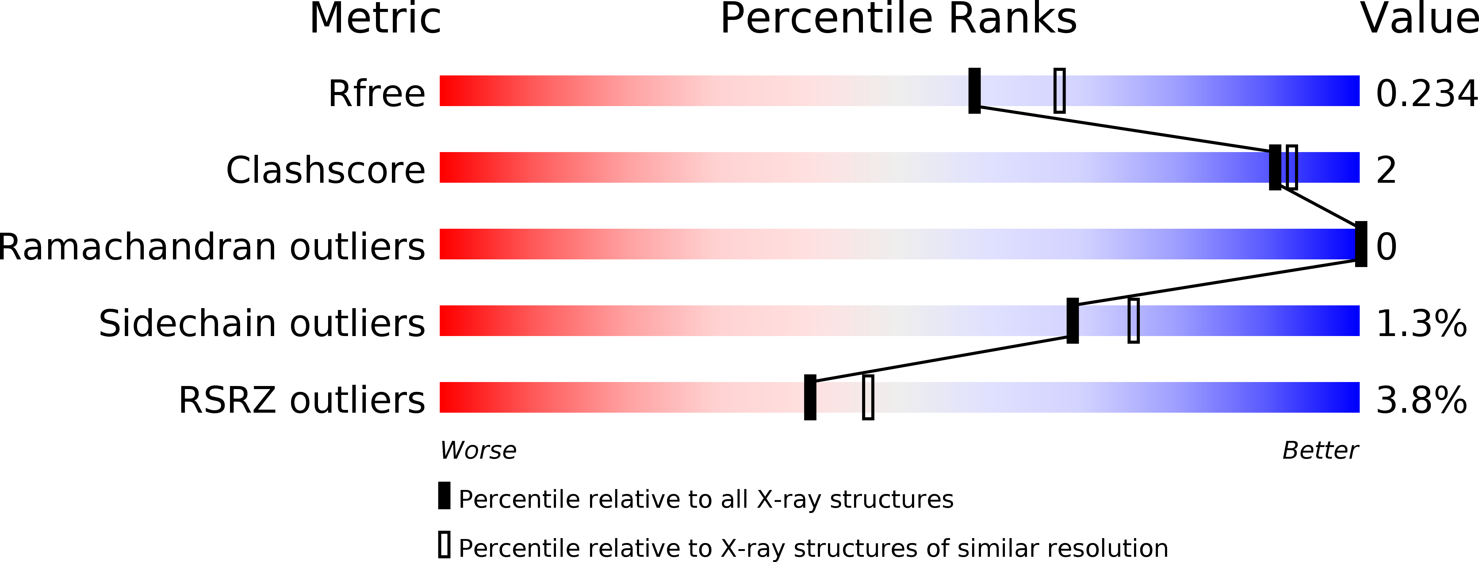
Resolution:
2.10 Å
R-Value Free:
0.22
R-Value Work:
0.17
R-Value Observed:
0.17
Space Group:
P 1 21 1
Macromolecular Entities
Structures with similar UniProt ID
Protein Blast
Polymer Type:polypeptide(L)
Molecule:Transthyretin-like protein
Gene (Uniprot):hiuH
Chain IDs:A, B, C, D
Chain Length:114
Number of Molecules:4
Biological Source:Escherichia coli
Primary Citation
The transthyretin-related protein: structural investigation of a novel protein family
J.Struct.Biol. 155 445 457 (2006)
PMID: 16723258 DOI: 10.1016/j.jsb.2006.04.002

Abstact

The transthyretin-related protein (TRP) family comprises proteins predicted to be structurally related to the homotetrameric transport protein transthyretin (TTR). The function of TRPs is not yet fully established, but recent data suggest that they are involved in purine catabolism. We have determined the three-dimensional structure of the Escherichia coli TRP in two crystal forms; one at 1.65 A resolution in the presence of zinc, and the other at 2.1 A resolution in the presence of zinc and bromide. The structures revealed five zinc-ion-binding sites per monomer. Of these, the zinc ions bound at sites I and II are coordinated in tetrahedral geometries to the side chains of residues His9, His96, His98, Ser114, and three water molecules at the putative ligand-binding site. Of these four residues, His9, His98, and Ser114 are conserved. His9 and His98 bind the central zinc (site I) together with two water molecules. The side chain of His98 also binds to the zinc ion at site II. Bromide ions bind at site I only, replacing one of the water molecules coordinated to the zinc ion. The C-terminal four amino acid sequence motif Y-[RK]-G-[ST] constitutes the signature sequence of the TRP family. Two Tyr111 residues form direct hydrogen bonds to each other over the tetramer interface at the area, which in TTR constitutes the rear part of its thyroxine-binding channel. The putative substrate/ligand-binding channel of TRP is consequently shallower and broader than its counterpart in TTR.

Legend

Protein

Chemical

Disease

Primary Citation of related structures