2exo image
Deposition Date 1994-07-11
Release Date 1995-02-07
Last Version Date 2024-10-23
Entry Detail
PDB ID:
2EXO
Title:
CRYSTAL STRUCTURE OF THE CATALYTIC DOMAIN OF THE BETA-1,4-GLYCANASE CEX FROM CELLULOMONAS FIMI
Biological Source:
Source Organism:
Method Details:
Experimental Method:
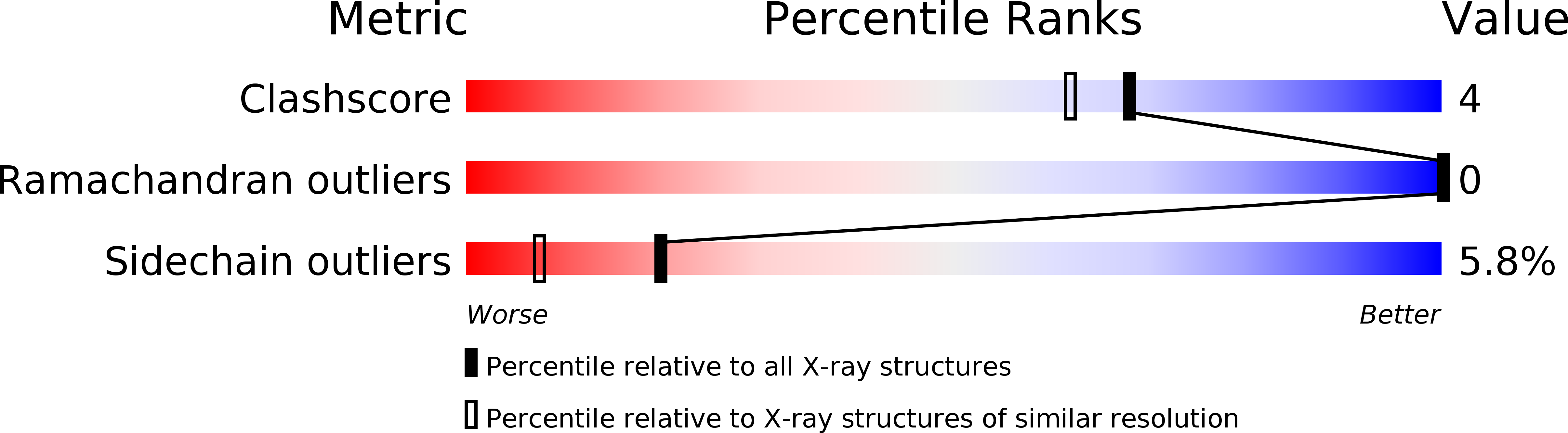
Resolution:
1.80 Å
R-Value Free:
0.26
R-Value Work:
0.21
R-Value Observed:
0.21
Space Group:
P 41 21 2
Macromolecular Entities
Structures with similar UniProt ID
Protein Blast
Polymer Type:polypeptide(L)
Molecule:EXO-1,4-BETA-D-GLYCANASE
Gene (Uniprot):cex
Chain IDs:A
Chain Length:312
Number of Molecules:1
Biological Source:Cellulomonas fimi
Primary Citation
Crystal structure of the catalytic domain of the beta-1,4-glycanase cex from Cellulomonas fimi.
Biochemistry 33 12546 12552 (1994)
PMID: 7918478 DOI: 10.1021/bi00208a003

Abstact

beta-1,4-Glycanases, principally cellulases and xylanases, are responsible for the hydrolysis of plant biomass. The bifunctional beta-1,4-xylanase/glucanase Cex from the bacterium Cellulomonas fimi, one of a large family of cellulases/xylanases, depolymerizes oligosaccharides and releases a disaccharide unit from the substrate nonreducing end. Hydrolysis occurs with net retention of the anomeric configuration of the sugar through a double-displacement mechanism involving a covalent glycosyl-enzyme intermediate. The active site nucleophile, Glu233, has been unambiguously identified by trapping of such an intermediate [Tull et al. (1991) J. Biol. Chem. 266, 15621-15625] and the acid/base catalyst, Glu127, by detailed kinetic analysis of mutants [MacLeod et al. (1994) Biochemistry 33, 6371-6376]. However, little is known about the enzyme's overall folding and its active site architecture. We report here the high-resolution crystal structure of the catalytic domain of Cex. The atomic structure refinement results in a model that includes 2400 protein atoms and 45 water molecules, with an R-factor of 0.217 for data extending to 1.8-A resolution. The protein forms an eight-parallel-stranded alpha/beta-barrel, which is a novel folding pattern for a microbial beta-glycanase. The active site, inferred from the location of Glu233, Glu127, and other conserved residues, is an open cleft on the carboxy-terminal end of the alpha/beta-barrel. An extensive hydrogen-bonding network stabilizes the ionization states of the key residues; in particular, the Asp235-His205-Glu233 hydrogen-bonding network may play a role in modulating the ionization state of Glu233 and in controlling local charge balance during the reaction.

Legend

Protein

Chemical

Disease

Primary Citation of related structures
Feedback Form
Name
Email
Institute
Feedback