2cw6 image
Deposition Date 2005-06-17
Release Date 2005-12-06
Last Version Date 2024-03-13
Entry Detail
PDB ID:
2CW6
Keywords:
Title:
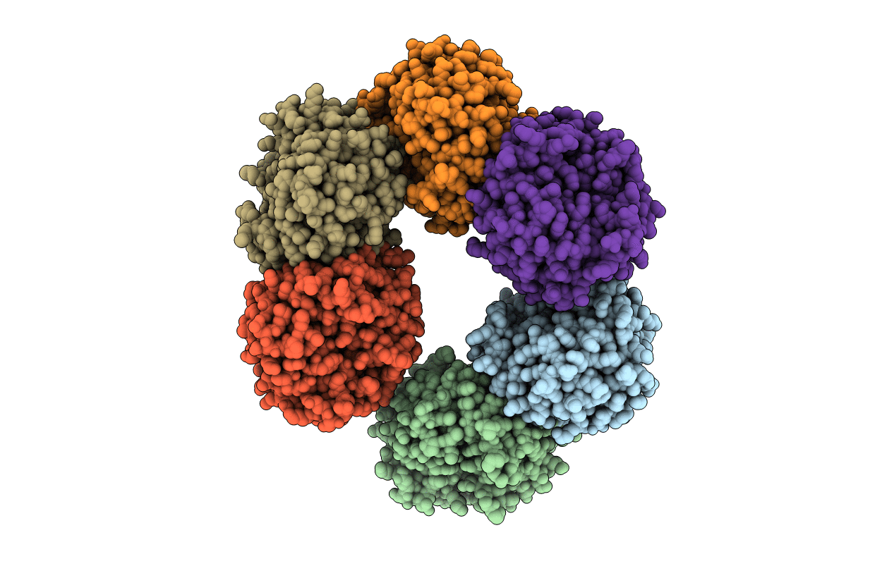
Crystal Structure of Human HMG-CoA Lyase: Insights into Catalysis and the Molecular Basis for Hydroxymethylglutaric Aciduria
Biological Source:
Source Organism:
Homo sapiens (Taxon ID: 9606)
Host Organism:
Method Details:
Experimental Method:
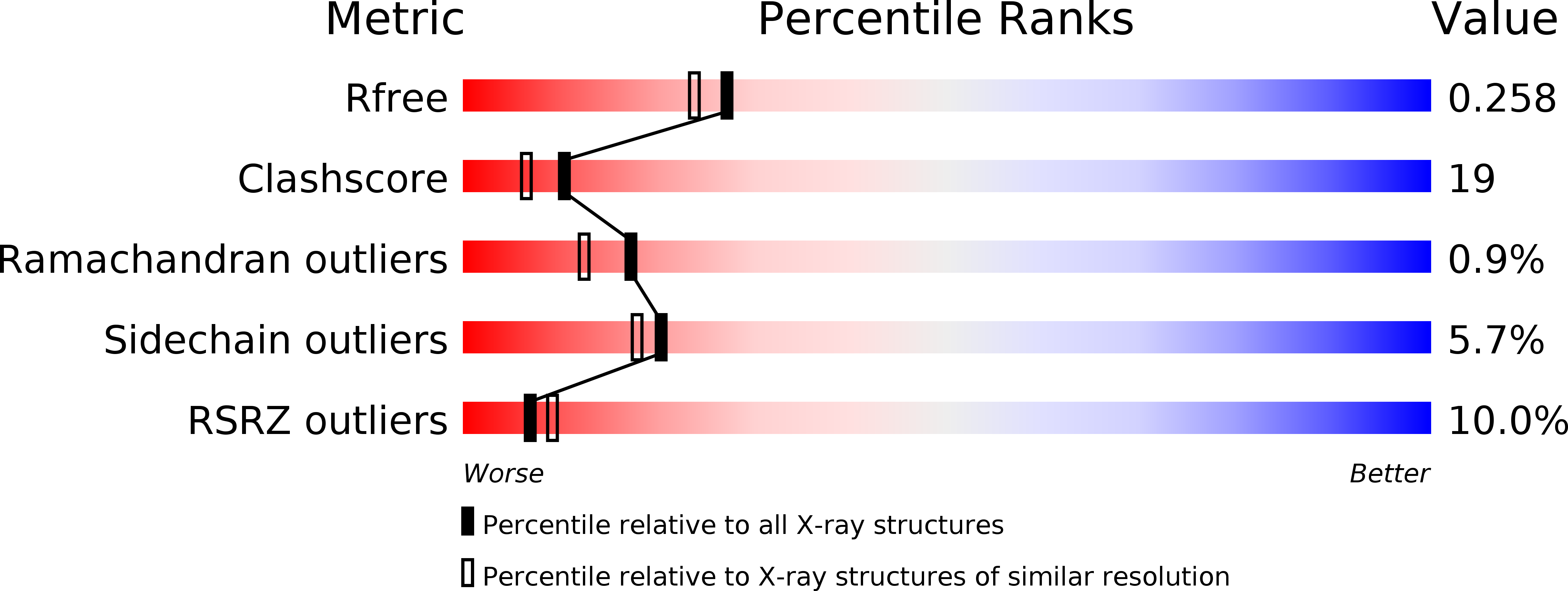
Resolution:
2.10 Å
R-Value Free:
0.26
R-Value Work:
0.22
R-Value Observed:
0.22
Space Group:
C 1 2 1
Macromolecular Entities
Structures with similar UniProt ID
Protein Blast
Polymer Type:polypeptide(L)
Molecule:Hydroxymethylglutaryl-CoA lyase, mitochondrial
Gene (Uniprot):HMGCL
Chain IDs:A, B, C, D, E, F
Chain Length:298
Number of Molecules:6
Biological Source:Homo sapiens
Primary Citation
Crystal structure of human 3-hydroxy-3-methylglutaryl-CoA Lyase: insights into catalysis and the molecular basis for hydroxymethylglutaric aciduria
J.Biol.Chem. 281 7526 7532 (2006)
PMID: 16330550 DOI: 10.1074/jbc.M506880200

Abstact

3-Hydroxy-3-methylglutaryl-CoA (HMG-CoA) lyase is a key enzyme in the ketogenic pathway that supplies metabolic fuel to extrahepatic tissues. Enzyme deficiency may be due to a variety of human mutations and can be fatal. Diminished activity has been explained based on analyses of recombinant human mutant proteins or, more recently, in the context of structural models for the enzyme. We report the experimental determination of a crystal structure at 2.1 A resolution of the recombinant human mitochondrial HMG-CoA lyase containing a bound activator cation and the dicarboxylic acid 3-hydroxyglutarate. The enzyme adopts a (betaalpha)(8) barrel fold, and the N-terminal barrel end is occluded. The structure of a physiologically relevant dimer suggests that substrate access to the active site involves binding across the cavity located at the C-terminal end of the barrel. An alternative hypothesis that involves substrate insertion through a pore proposed to extend through the barrel is not compatible with the observed structure. The activator cation ligands included Asn(275), Asp(42),His(233), and His(235); the latter three residues had been implicated previously as contributing to metal binding or enzyme activity. Arg(41), previously shown to have a major effect on catalytic efficiency, is also located at the active site. In the observed structure, this residue interacts with a carboxyl group of 3-hydroxyglutarate, the hydrolysis product of the competitive inhibitor 3-hydroxyglutaryl-CoA required for crystallization of human enzyme. The structure provides a rationale for the decrease in enzyme activity due to clinical mutations, including H233R, R41Q, D42H, and D204N, that compromise active site function or enzyme stability.

Legend

Protein

Chemical

Disease

Primary Citation of related structures