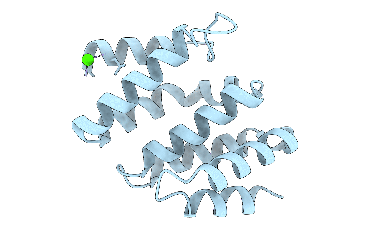
2bf0 image
Deposition Date 2004-12-02
Release Date 2005-01-18
Last Version Date 2024-11-06
Entry Detail
PDB ID:
2BF0
Keywords:
Title:
crystal structure of the rpr of pcf11
Biological Source:
Source Organism:
Host Organism:
Method Details:
Experimental Method:
Resolution:
2.30 Å
R-Value Free:
0.28
R-Value Work:
0.25
R-Value Observed:
0.26
Space Group:
P 43 21 2
Macromolecular Entities
Structures with similar UniProt ID
Protein Blast
Polymer Type:polypeptide(L)
Molecule:PCF11
Gene (Uniprot):PCF11
Chain IDs:A (auth: X)
Chain Length:143
Number of Molecules:1
Biological Source:SACCHAROMYCES CEREVISIAE
Modified Residue
Compound ID Chain ID Parent Comp ID Details 2D Image
MSE A MET SELENOMETHIONINE
Ligand Molecules
Primary Citation
Key Features of the Interaction between Pcf11 Cid and RNA Polymerase II Ctd.
Nat.Struct.Mol.Biol. 12 144 ? (2005)
PMID: 15665873 DOI: 10.1038/NSMB887

Abstact

The C-terminal domain (CTD) of the large subunit of RNA polymerase II is a platform for mRNA processing factors and links gene transcription to mRNA capping, splicing and polyadenylation. Pcf11, an essential component of the mRNA cleavage factor IA, contains a CTD-interaction domain that binds in a phospho-dependent manner to the heptad repeats within the RNA polymerase II CTD. We show here that the phosphorylated CTD exists as a dynamic disordered ensemble in solution and, by induced fit, it assumes a structured conformation when bound to Pcf11. In addition, we detected cis-trans populations for the CTD prolines, and found that only the all-trans form is selected for binding. These data suggest that the recognition of the CTD is regulated by independent site-specific modifications (phosphorylation and proline cis-trans isomerization) and, probably, by the local concentration of suitable binding sites.

Legend

Protein

Chemical

Disease

Primary Citation of related structures
Feedback Form
Name
Email
Institute
Feedback