2bc0 image
Deposition Date 2005-10-18
Release Date 2006-10-31
Last Version Date 2024-10-30
Entry Detail
PDB ID:
2BC0
Keywords:
Title:
Structural Analysis of Streptococcus pyogenes NADH oxidase: Wild-type Nox
Biological Source:
Source Organism:
Host Organism:
Method Details:
Experimental Method:
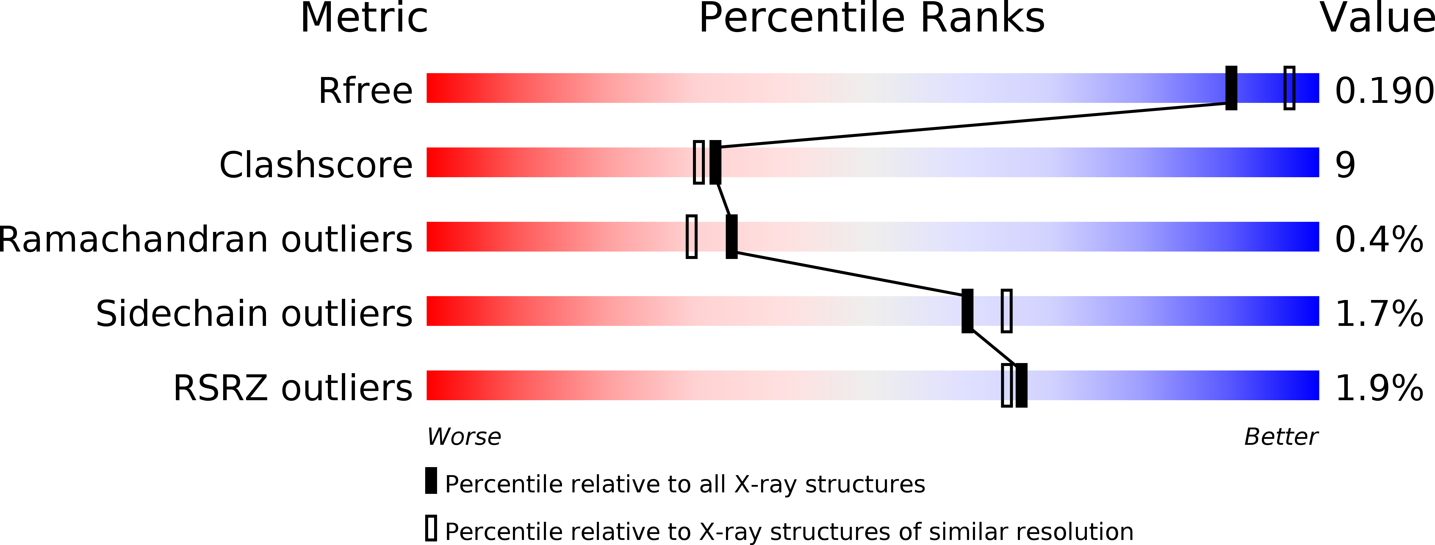
Resolution:
2.00 Å
R-Value Free:
0.23
R-Value Work:
0.19
R-Value Observed:
0.19
Space Group:
P 21 21 21
Macromolecular Entities
Polymer Type:polypeptide(L)
Molecule:NADH Oxidase
Chain IDs:A, B
Chain Length:490
Number of Molecules:2
Biological Source:Streptococcus pyogenes
Modified Residue
Compound ID Chain ID Parent Comp ID Details 2D Image
CSD A CYS 3-SULFINOALANINE
Ligand Molecules
Primary Citation
Structural Analysis of Streptococcus pyogenes NADH Oxidase: Conformational Dynamics Involved in Formation of the C(4a)-Peroxyflavin Intermediate.
Biochemistry 54 6815 6829 (2015)
PMID: 26506002 DOI: 10.1021/acs.biochem.5b00676

Abstact

In probing the oxygen reactivity of an Enterococcus faecalis NADH oxidase (Nox; O2 → 2H2O) C42S mutant lacking the Cys42-sulfenic acid (Cys42-SOH) redox center, we provided direct evidence of a C(4a)-peroxyflavin intermediate in the oxidative half-reaction and also described a conformational or chemical change that is rate-limiting for full reoxidation of the homodimer. In this work, the Nox from Streptococcus pyogenes (SpyNox) has been expressed and crystallized, and the overoxidized wild-type [Cys44-SOH → Cys44-sulfinic acid (Cys44-SO2H)] and C44S mutant enzyme structures have been refined at 2.0 and 2.15 Å, respectively. We show that azide binds to the two-electron reduced wild-type (EH2) enzyme and to the mutant enzyme in solution, but with a significantly higher affinity for the mutant protein. The spectral course of the titration with the SpyNox EH2 form clearly indicates progressive displacement of the Cys44-S(-) → FAD charge-transfer interaction. An azide soak with C44S Nox crystals led to the structure of the complex, as refined at 2.10 Å. The active-site N3(-) ligand is proximal to the Ser44 and His11 side chains, and a significant shift in the Ser44 side chain also appears. This provides an attractive explanation for the azide-induced loss of charge-transfer absorbance seen with the wild-type EH2 form and also permits accommodation of a C(4a)-peroxyflavin structural model. The conformation of Ser44 and the associated helical element, and the resulting steric accommodation, appear to be linked to the conformational change described in the E. faecalis C42S Nox oxidative half-reaction.

Legend

Protein

Chemical

Disease

Primary Citation of related structures