Deposition Date
2009-02-18
Release Date
2009-10-06
Last Version Date
2023-11-01
Entry Detail
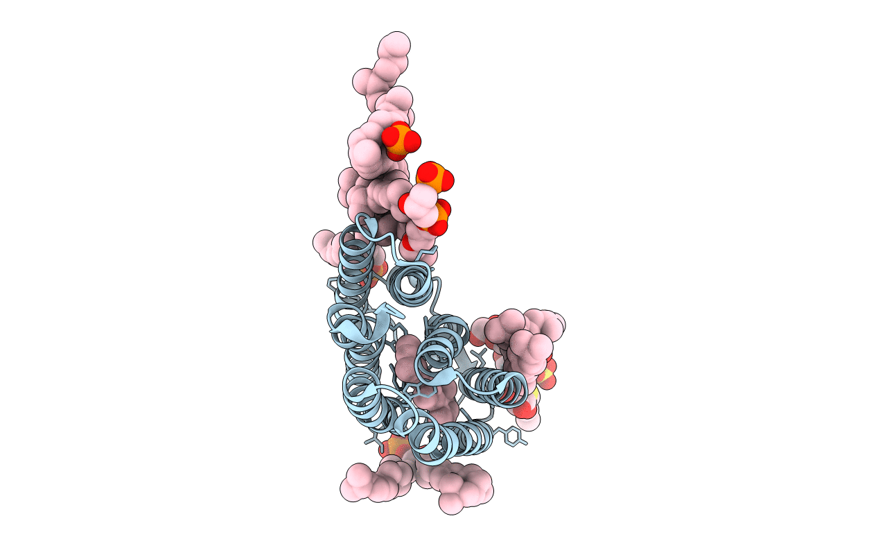
PDB ID:
2ZZL
Keywords:
Title:
Structure of bacteriorhodopsin's M intermediate at pH 7
Biological Source:
Source Organism(s):
Halobacterium salinarum (Taxon ID: 2242)
Method Details:
Experimental Method:
Resolution:
2.03 Å
R-Value Free:
0.23
R-Value Work:
0.20
R-Value Observed:
0.20
Space Group:
P 6 2 2