2ZZJ image
Deposition Date 2009-02-16
Release Date 2009-05-05
Last Version Date 2024-11-13
Entry Detail
PDB ID:
2ZZJ
Keywords:
Title:
Crystal structure of endo-beta-1,4-glucuronan lyase from fungus Trichoderma reesei
Biological Source:
Source Organism:
Host Organism:
Method Details:
Experimental Method:
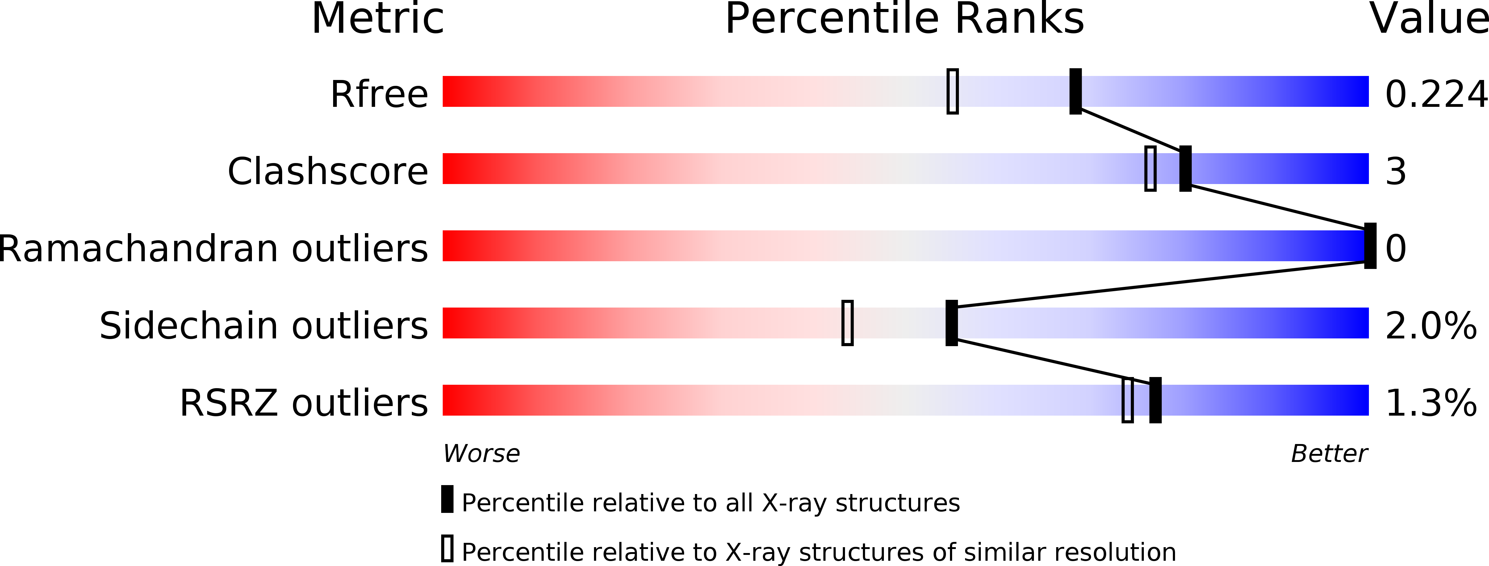
Resolution:
1.80 Å
R-Value Free:
0.22
R-Value Work:
0.18
R-Value Observed:
0.18
Space Group:
P 21 21 21
Macromolecular Entities
Structures with similar UniProt ID
Protein Blast
Polymer Type:polypeptide(L)
Molecule:Glucuronan lyase A
Gene (Uniprot):glA
Chain IDs:A
Chain Length:238
Number of Molecules:1
Biological Source:Trichoderma reesei
Primary Citation
Crystal structure of polysaccharide lyase family 20 endo-beta-1,4-glucuronan lyase from the filamentous fungus Trichoderma reesei.
Febs Lett. 583 1323 1326 (2009)
PMID: 19306878 DOI: 10.1016/j.febslet.2009.03.034

Abstact

The crystal structure of endo-beta-(1-->4)-glucuronan lyase from Trichoderma reesei (TrGL) has been determined at 1.8A resolution as the first three-dimensional structure of polysaccharide lyase (PL) family 20. TrGL has a typical beta-jelly roll fold, which is similar to glycoside hydrolase family 16 and PL7 enzymes. A calcium ion is bound to the site far from the cleft and appears to contribute to the stability. There are several completely conserved residues in the cleft. Possible catalytic residues are predicted based on structural comparison with PL7 alginate lyase A1-II'.

Legend

Protein

Chemical

Disease

Primary Citation of related structures