2ZYO image
Deposition Date 2009-01-27
Release Date 2009-03-31
Last Version Date 2023-11-01
Entry Detail
PDB ID:
2ZYO
Title:
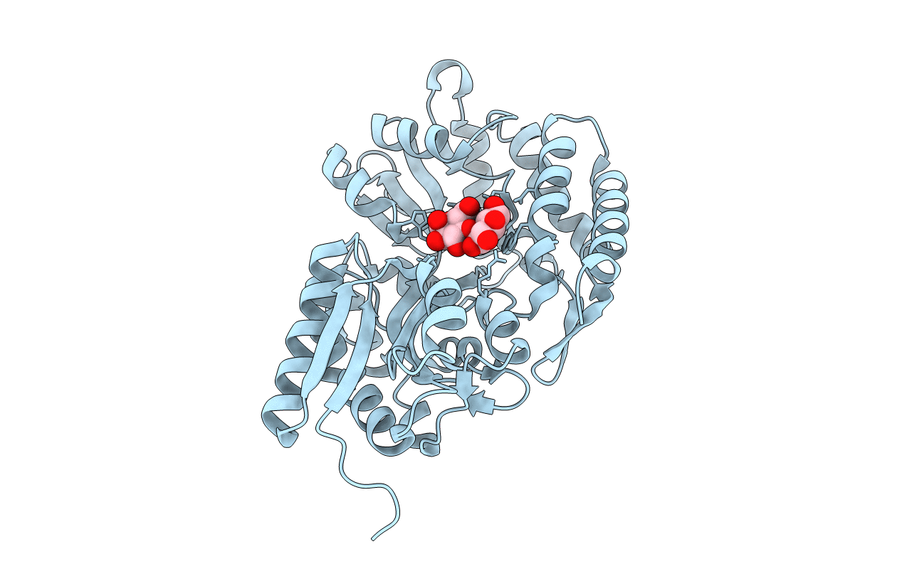
Crystal structure of cyclo/maltodextrin-binding protein complexed with maltotetraose
Biological Source:
Source Organism:
Host Organism:
Method Details:
Experimental Method:
Resolution:
1.55 Å
R-Value Free:
0.22
R-Value Work:
0.19
R-Value Observed:
0.19
Space Group:
P 21 21 21
Macromolecular Entities
Structures with similar UniProt ID
Protein Blast
Polymer Type:polypeptide(L)
Molecule:solute-binding protein
Gene (Uniprot):orf-3
Chain IDs:A
Chain Length:397
Number of Molecules:1
Biological Source:Thermoactinomyces vulgaris
Peptide-like Molecules
PRD_900001
Primary Citation
Crystal structures of open and closed forms of cyclo/maltodextrin-binding protein
Febs J. 276 3008 3019 (2009)
PMID: 19490104 DOI: 10.1111/j.1742-4658.2009.07020.x

Abstact

The crystal structures of Thermoactinomyces vulgaris cyclo/maltodextrin-binding protein (TvuCMBP) complexed with alpha-cyclodextrin (alpha-CD), beta-cyclodextrin (beta-CD) and maltotetraose (G4) have been determined. A common functional conformational change among all solute-binding proteins involves switching from an open form to a closed form, which facilitates transporter binding. Escherichia coli maltodextrin-binding protein (EcoMBP), which is structurally homologous to TvuCMBP, has been determined to adopt the open form when complexed with beta-CD and the closed form when bound to G4. Here, we show that, unlike EcoMBP, TvuCMBP-alpha-CD and TvuCMBP-beta-CD adopt the closed form when complexed, whereas TvuCMBP-G4 adopts the open form. Only two glucose residues are evident in the TvuCMBP-G4 structure, and these bind to the C-domain of TvuCMBP in a manner similar to the way in which maltose binds to the C-domain of EcoMBP. The superposition of TvuCMBP-alpha-CD, TvuCMBP-beta-CD and TvuCMBP-gamma-CD shows that the positions and the orientations of three glucose residues in the cyclodextrin molecules overlay remarkably well. In addition, most of the amino acid residues interacting with these three glucose residues also participate in interactions with the two glucose residues in TvuCMBP-G4, regardless of whether the protein is in the closed or open form. Our results suggest that the mechanisms by which TvuCMBP changes from the open to the closed conformation and maintains the closed form appear to be different from those of EcoMBP, despite the fact that the amino acid residues responsible for the initial binding of the ligands are well conserved between TvuCMBP and EcoMBP.

Legend

Protein

Chemical

Disease

Primary Citation of related structures
Feedback Form
Name
Email
Institute
Feedback