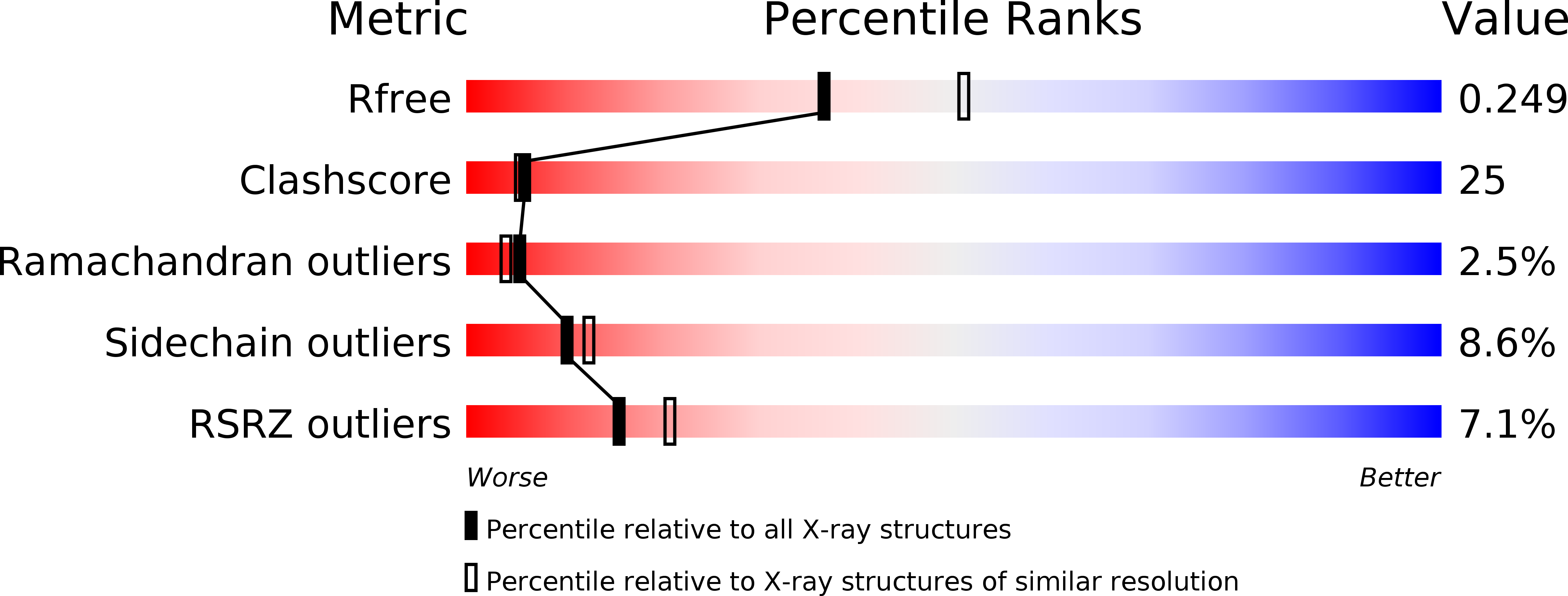
2ZXP image
Deposition Date 2009-01-05
Release Date 2010-01-05
Last Version Date 2023-11-01
Entry Detail
PDB ID:
2ZXP
Keywords:
Title:
Crystal structure of RecJ in complex with Mn2+ from Thermus thermophilus HB8
Biological Source:
Source Organism:
Host Organism:
Method Details:
Experimental Method:
Resolution:
2.30 Å
R-Value Free:
0.28
R-Value Work:
0.23
Space Group:
P 43 21 2
Macromolecular Entities
Polymer Type:polypeptide(L)
Molecule:Single-stranded DNA specific exonuclease RecJ
Gene (Uniprot):recJ
Chain IDs:A
Chain Length:666
Number of Molecules:1
Biological Source:Thermus thermophilus
Ligand Molecules
Primary Citation
Structure of RecJ exonuclease defines its specificity for single-stranded DNA
J.Biol.Chem. 285 9762 9769 (2010)
PMID: 20129927 DOI: 10.1074/jbc.M109.096487

Abstact

RecJ is a single-stranded DNA (ssDNA)-specific 5'-3' exonuclease that plays an important role in DNA repair and recombination. To elucidate how RecJ achieves its high specificity for ssDNA, we determined the entire structures of RecJ both in a ligand-free form and in a complex with Mn(2+) or Mg(2+) by x-ray crystallography. The entire RecJ consists of four domains that form a molecule with an O-like structure. One of two newly identified domains had structural similarities to an oligonucleotide/oligosaccharide-binding (OB) fold. The OB fold domain alone could bind to DNA, indicating that this domain is a novel member of the OB fold superfamily. The truncated RecJ containing only the core domain exhibited much lower affinity for the ssDNA substrate compared with intact RecJ. These results support the hypothesis that these structural features allow specific binding of RecJ to ssDNA. In addition, the structure of the RecJ-Mn(2+) complex suggests that the hydrolysis reaction catalyzed by RecJ proceeds through a two-metal ion mechanism.

Legend

Protein

Chemical

Disease

Primary Citation of related structures