2ZXI image
Deposition Date 2008-12-24
Release Date 2009-05-19
Last Version Date 2023-11-01
Entry Detail
PDB ID:
2ZXI
Title:
Structure of Aquifex aeolicus GidA in the form II crystal
Biological Source:
Source Organism:
Method Details:
Experimental Method:
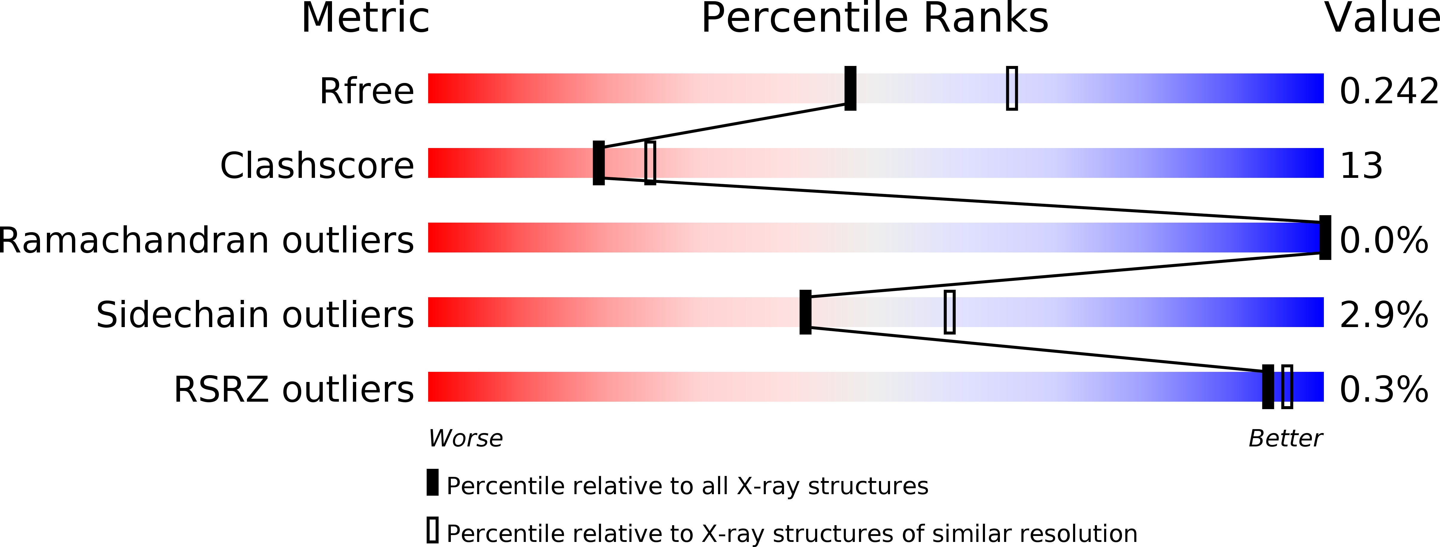
Resolution:
2.30 Å
R-Value Free:
0.24
R-Value Work:
0.20
R-Value Observed:
0.20
Space Group:
P 1 21 1
Macromolecular Entities
Structures with similar UniProt ID
Protein Blast
Polymer Type:polypeptide(L)
Molecule:tRNA uridine 5-carboxymethylaminomethyl modification enzyme mnmG
Gene (Uniprot):mnmG
Chain IDs:A, B, C, D
Chain Length:637
Number of Molecules:4
Biological Source:Aquifex aeolicus
Ligand Molecules
Primary Citation
Conserved cysteine residues of GidA are essential for biogenesis of 5-carboxymethylaminomethyluridine at tRNA anticodon
Structure 17 713 724 (2009)
PMID: 19446527 DOI: 10.1016/j.str.2009.03.013

Abstact

The 5-carboxymethylaminomethyl modification of uridine (cmnm(5)U) at the anticodon first position occurs in tRNAs that read split codon boxes ending with purine. This modification is crucial for correct translation, by restricting codon-anticodon wobbling. Two conserved enzymes, GidA and MnmE, participate in the cmnm(5)U modification process. Here we determined the crystal structure of Aquifex aeolicus GidA at 2.3 A resolution. The structure revealed the tight interaction of GidA with FAD. Structure-based mutation analyses allowed us to identify two conserved Cys residues in the vicinity of the FAD-binding site that are essential for the cmnm(5)U modification in vivo. Together with mutational analysis of MnmE, we propose a mechanism for the cmnm(5)U modification process where GidA, but not MnmE, attacks the C6 atom of uridine by a mechanism analogous to that of thymidylate synthase. We also present a tRNA-docking model that provides structural insights into the tRNA recognition mechanism for efficient modification.

Legend

Protein

Chemical

Disease

Primary Citation of related structures
Feedback Form
Name
Email
Institute
Feedback