2ZXE image
Deposition Date 2008-12-22
Release Date 2009-05-19
Last Version Date 2024-11-06
Entry Detail
PDB ID:
2ZXE
Title:
Crystal structure of the sodium - potassium pump in the E2.2K+.Pi state
Biological Source:
Source Organism:
Method Details:
Experimental Method:
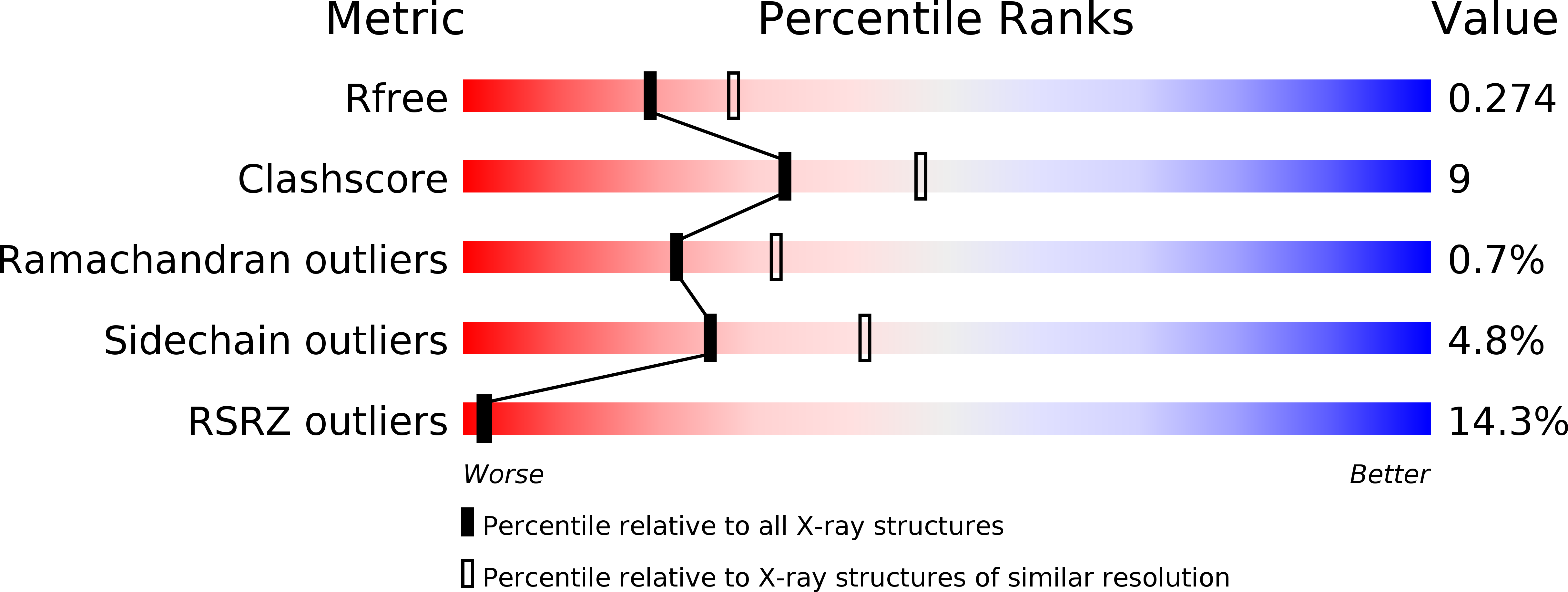
Resolution:
2.40 Å
R-Value Free:
0.27
R-Value Work:
0.24
R-Value Observed:
0.24
Space Group:
C 1 2 1
Macromolecular Entities
Polymer Type:polypeptide(L)
Molecule:Na, K-ATPase alpha subunit
Gene (Uniprot):atn1
Chain IDs:A
Chain Length:1028
Number of Molecules:1
Biological Source:Squalus acanthias
Polymer Type:polypeptide(L)
Molecule:Na+,K+-ATPase beta subunit
Chain IDs:B
Chain Length:305
Number of Molecules:1
Biological Source:Squalus acanthias
Polymer Type:polypeptide(L)
Molecule:Phospholemman-like protein
Gene (Uniprot):fxyd10
Chain IDs:C (auth: G)
Chain Length:74
Number of Molecules:1
Biological Source:Squalus acanthias
Modified Residue
Compound ID Chain ID Parent Comp ID Details 2D Image
ASN B ASN GLYCOSYLATION SITE
Primary Citation
Crystal structure of the sodium - potassium pump at 2.4 A resolution
Nature 459 446 450 (2009)
PMID: 19458722 DOI: 10.1038/nature07939

Abstact

Sodium-potassium ATPase is an ATP-powered ion pump that establishes concentration gradients for Na(+) and K(+) ions across the plasma membrane in all animal cells by pumping Na(+) from the cytoplasm and K(+) from the extracellular medium. Such gradients are used in many essential processes, notably for generating action potentials. Na(+), K(+)-ATPase is a member of the P-type ATPases, which include sarcoplasmic reticulum Ca(2+)-ATPase and gastric H(+), K(+)-ATPase, among others, and is the target of cardiac glycosides. Here we describe a crystal structure of this important ion pump, from shark rectal glands, consisting of alpha- and beta-subunits and a regulatory FXYD protein, all of which are highly homologous to human ones. The ATPase was fixed in a state analogous to E2.2K(+).P(i), in which the ATPase has a high affinity for K(+) and still binds P(i), as in the first crystal structure of pig kidney enzyme at 3.5 A resolution. Clearly visualized now at 2.4 A resolution are coordination of K(+) and associated water molecules in the transmembrane binding sites and a phosphate analogue (MgF(4)(2-)) in the phosphorylation site. The crystal structure shows that the beta-subunit has a critical role in K(+) binding (although its involvement has previously been suggested) and explains, at least partially, why the homologous Ca(2+)-ATPase counter-transports H(+) rather than K(+), despite the coordinating residues being almost identical.

Legend

Protein

Chemical

Disease

Primary Citation of related structures