
Deposition Date
2008-11-14
Release Date
2009-03-10
Last Version Date
2024-03-13
Entry Detail
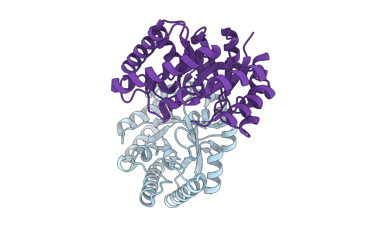
PDB ID:
2ZVR
Keywords:
Title:
Crystal structure of a D-tagatose 3-epimerase-related protein from Thermotoga maritima
Biological Source:
Source Organism(s):
Thermotoga maritima (Taxon ID: 2336)
Expression System(s):
Method Details:
Experimental Method:
Resolution:
2.20 Å
R-Value Free:
0.24
R-Value Work:
0.19
R-Value Observed:
0.19
Space Group:
P 1
