2ZUQ image
Deposition Date 2008-10-28
Release Date 2009-04-14
Last Version Date 2024-10-23
Entry Detail
PDB ID:
2ZUQ
Title:
Crystal structure of DsbB-Fab complex
Biological Source:
Source Organism:
Escherichia coli (Taxon ID: 83333)
mus musculus (Taxon ID: 10090)
Host Organism:
Method Details:
Experimental Method:
Resolution:
3.30 Å
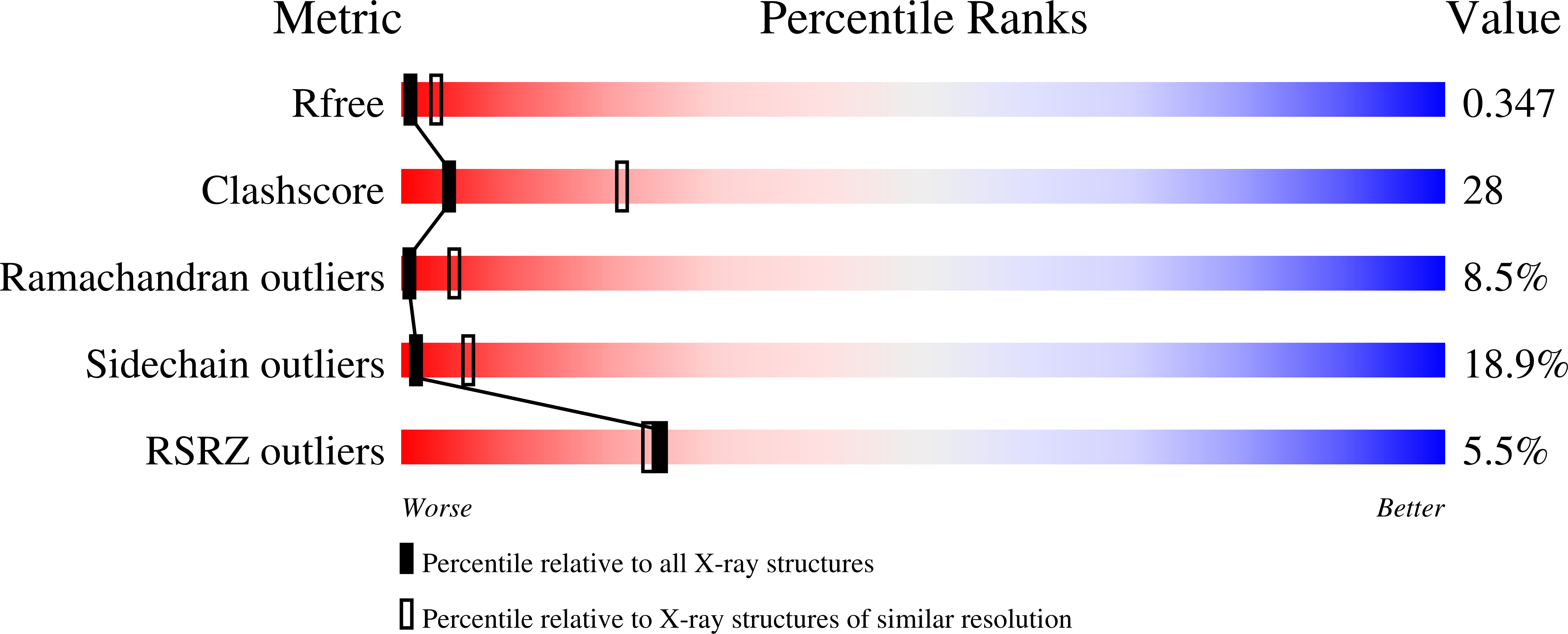
R-Value Free:
0.35
R-Value Work:
0.27
R-Value Observed:
0.27
Space Group:
C 1 2 1
Macromolecular Entities
Polymer Type:polypeptide(L)
Molecule:Disulfide bond formation protein B
Gene (Uniprot):dsbB
Mutations:C8A,C41S,C49V
Chain IDs:A, D
Chain Length:176
Number of Molecules:2
Biological Source:Escherichia coli
Polymer Type:polypeptide(L)
Molecule:Fab fragment light chain
Chain IDs:B, E
Chain Length:239
Number of Molecules:2
Biological Source:mus musculus
Polymer Type:polypeptide(L)
Molecule:Fab fragment heavy chain
Chain IDs:C, F
Chain Length:221
Number of Molecules:2
Biological Source:mus musculus
Ligand Molecules
Primary Citation
Dynamic nature of disulphide bond formation catalysts revealed by crystal structures of DsbB
Embo J. 28 779 791 (2009)
PMID: 19214188 DOI: 10.1038/emboj.2009.21

Abstact

In the Escherichia coli system catalysing oxidative protein folding, disulphide bonds are generated by the cooperation of DsbB and ubiquinone and transferred to substrate proteins through DsbA. The structures solved so far for different forms of DsbB lack the Cys104-Cys130 initial-state disulphide that is directly donated to DsbA. Here, we report the 3.4 A crystal structure of a DsbB-Fab complex, in which DsbB has this principal disulphide. Its comparison with the updated structure of the DsbB-DsbA complex as well as with the recently reported NMR structure of a DsbB variant having the rearranged Cys41-Cys130 disulphide illuminated conformational transitions of DsbB induced by the binding and release of DsbA. Mutational studies revealed that the membrane-parallel short alpha-helix of DsbB has a key function in physiological electron flow, presumably by controlling the positioning of the Cys130-containing loop. These findings demonstrate that DsbB has developed the elaborate conformational dynamism to oxidize DsbA for continuous protein disulphide bond formation in the cell.

Legend

Protein

Chemical

Disease

Primary Citation of related structures