Deposition Date
2008-10-15
Release Date
2009-04-07
Last Version Date
2023-11-01
Entry Detail
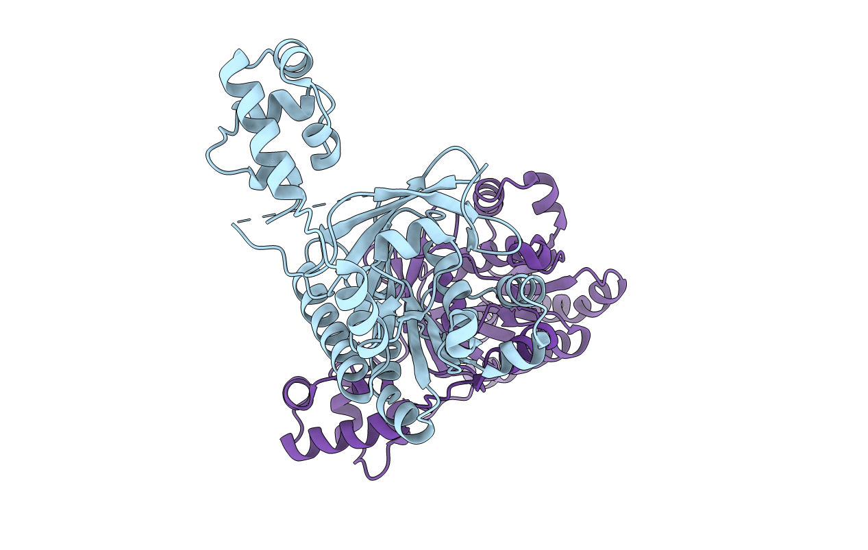
Biological Source:
Source Organism:
Sulfolobus solfataricus (Taxon ID: 2287)
Host Organism:
Method Details:
Experimental Method:
Resolution:
3.30 Å
R-Value Free:
0.28
R-Value Work:
0.22
R-Value Observed:
0.23
Space Group:
P 21 21 21