
Deposition Date
2008-10-12
Release Date
2009-01-13
Last Version Date
2023-11-01
Entry Detail
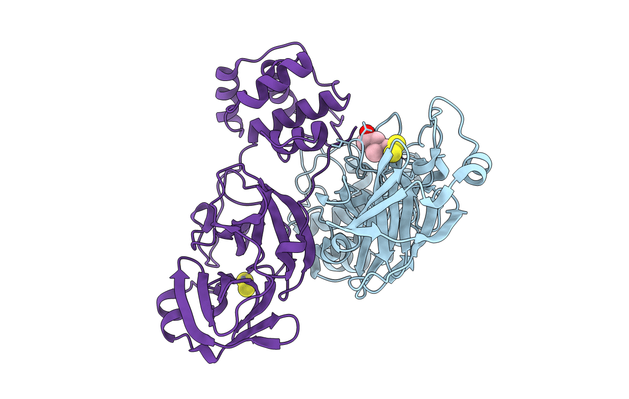
PDB ID:
2ZU2
Keywords:
Title:
complex structure of CoV 229E 3CL protease with EPDTC
Biological Source:
Source Organism(s):
Human coronavirus (Taxon ID: 11137)
Expression System(s):
Method Details:
Experimental Method:
Resolution:
1.80 Å
R-Value Free:
0.22
R-Value Work:
0.18
R-Value Observed:
0.19
Space Group:
P 1 21 1
