2ZU0 image
Deposition Date 2008-10-11
Release Date 2009-03-10
Last Version Date 2023-11-01
Entry Detail
PDB ID:
2ZU0
Title:
Crystal structure of SufC-SufD complex involved in the iron-sulfur cluster biosynthesis
Biological Source:
Source Organism:
Host Organism:
Method Details:
Experimental Method:
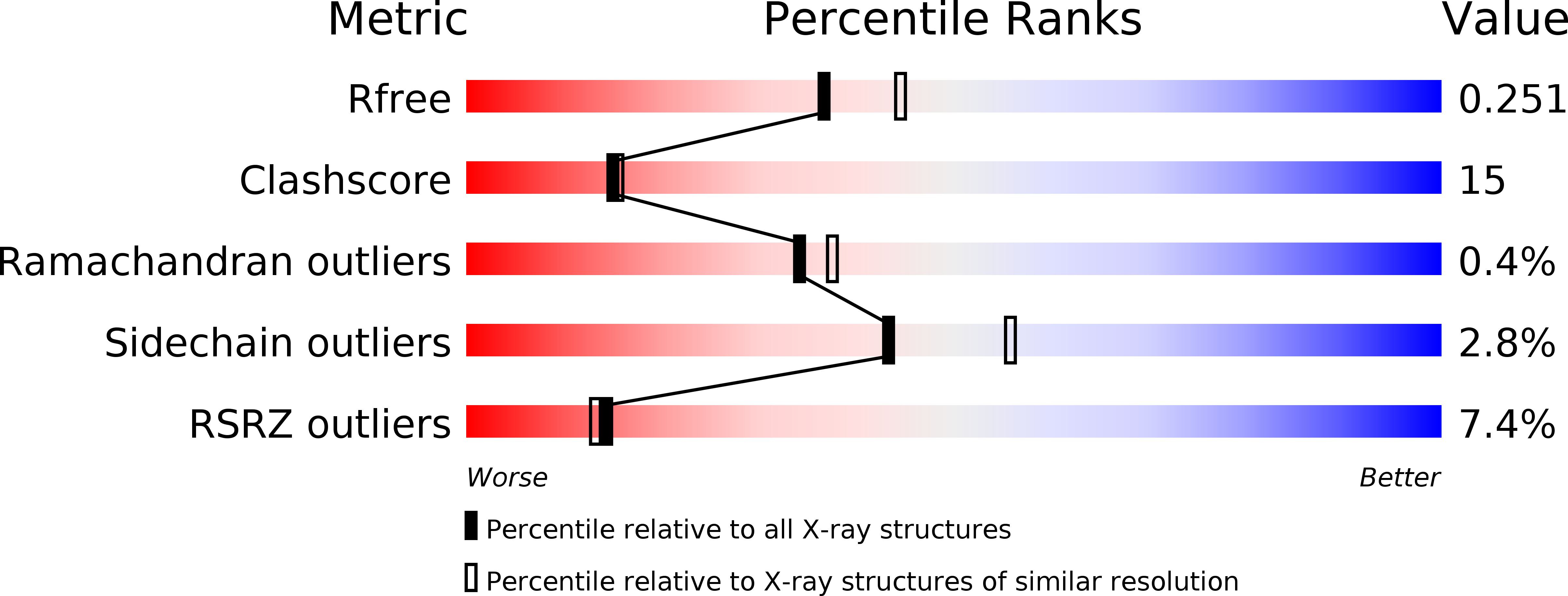
Resolution:
2.20 Å
R-Value Free:
0.25
R-Value Work:
0.23
R-Value Observed:
0.23
Space Group:
P 21 21 21
Macromolecular Entities
Structures with similar UniProt ID
Protein Blast
Polymer Type:polypeptide(L)
Molecule:Protein sufD
Gene (Uniprot):sufD
Chain IDs:A, B
Chain Length:423
Number of Molecules:2
Biological Source:Escherichia coli
Structures with similar UniProt ID
Protein Blast
Polymer Type:polypeptide(L)
Molecule:Probable ATP-dependent transporter sufC
Gene (Uniprot):sufC
Chain IDs:C, D
Chain Length:267
Number of Molecules:2
Biological Source:Escherichia coli
Ligand Molecules
Primary Citation
Molecular dynamism of Fe-S cluster biosynthesis implicated by the structure of SufC(2)-SufD(2) complex
J.Mol.Biol. 387 245 258 (2009)
PMID: 19361433 DOI: 10.1016/j.jmb.2009.01.054

Abstact

Maturation of iron-sulfur (Fe-S) proteins is achieved by the SUF machinery in a wide number of eubacteria and archaea, as well as eukaryotic chloroplasts. This machinery is encoded in Escherichia coli by the sufABCDSE operon, where three Suf components, SufB, SufC, and SufD, form a complex and appear to provide an intermediary site for the Fe-S cluster assembly. Here, we report the quaternary structure of the SufC(2)-SufD(2) complex in which SufC is bound to the C-terminal domain of SufD. Comparison with the monomeric structure of SufC revealed conformational change of the active-site residues: SufC becomes competent for ATP binding and hydrolysis upon association with SufD. The two SufC subunits were spatially separated in the SufC(2)-SufD(2) complex, whereas cross-linking experiments in solution have indicated that two SufC molecules associate with each other in the presence of Mg(2+) and ATP. Such dimer formation of SufC may lead to a gross structural change of the SufC(2)-SufD(2) complex. Furthermore, genetic analysis of SufD revealed an essential histidine residue buried inside the dimer interface, suggesting that conformational change may expose this crucial residue. These findings, together with biochemical characterization of the SufB-SufC-SufD complex, have led us to propose a model for the Fe-S cluster biosynthesis in the complex.

Legend

Protein

Chemical

Disease

Primary Citation of related structures
Feedback Form
Name
Email
Institute
Feedback